Vardaan Learning Institute
Secondary Course • Science and Technology (212)
Chapter 22: Life Processes: Nutrition, Respiration & Transport
Part I: Nutrition • Part II: Respiration • Part III: Transportation • Part IV: Excretion
1. Fundamentals of Life Processes
All living organisms exhibit specialized characteristics that distinguish them from non-living matter. Even while resting, asleep, or sitting quietly, continuous maintenance jobs must occur inside cells to prevent damage and destruction of cellular architecture.
Core Definition
Life Processes are the fundamental biological activities and maintenance functions performed by living organisms to maintain life, repair cellular structures, sustain metabolic balance (homeostasis), and ensure survival on Earth.
The four primary vital life processes essential for every living organism are:
- Nutrition: The intake and biochemical conversion of food/nutrients to supply chemical energy and raw materials for cellular maintenance, biosynthesis, and repair.
- Respiration: The step-by-step biochemical oxidation and catabolism of food molecules (chiefly glucose) inside cells to generate usable metabolic energy stored in the form of Adenosine Triphosphate (ATP).
- Transportation: The internal translocation mechanism responsible for moving nutrients, respiratory gases ($\mathrm{O_2}$, $\mathrm{CO_2}$), water, minerals, and metabolic wastes between different specialized organs and cells.
- Excretion: The physiological process of eliminating potentially toxic nitrogenous metabolic end-products (such as urea, uric acid, and excess salts) from the internal environment.
Living organisms are highly organized structures made of tissues, tissues of cells, and cells of smaller molecular assemblies. Environmental influences constantly tend to break down this ordered structure over time. If order breaks down, the organism cannot stay alive. Therefore, living creatures must continuously move molecules around internally to repair, maintain, and sustain cellular integrity.
2. Nutrition: Modes and Mechanisms
Every organism requires energy to perform work and carbon-based molecules to grow, heal, and manufacture enzymes and structural proteins. Organisms fulfill these demands through different modes of nutrition:
| Feature |
Autotrophic Nutrition |
Heterotrophic Nutrition |
| Source of Carbon |
Inorganic substances: Carbon dioxide ($\mathrm{CO_2}$) and Water ($\mathrm{H_2O}$). |
Complex organic substances derived directly or indirectly from other organisms. |
| Energy Input |
Solar radiant energy (captured by chlorophyll) or chemical energy. |
Chemical bond energy contained in ingested organic food. |
| Chlorophyll |
Mandatory for photosynthetic autotrophs. |
Absent. |
| Representative Organisms |
Green plants, Euglena, Cyanobacteria (blue-green algae). |
Animals, Fungi (moulds, mushrooms), Protozoans, majority of Bacteria. |
A. Autotrophic Nutrition (Photosynthesis)
Photosynthesis is the foundational photochemical process by which green plants manufacture carbohydrates (glucose) from carbon dioxide and water in the presence of sunlight and chlorophyll, releasing oxygen as an essential byproduct.
Three Sequential Events Occurring During Photosynthesis:
- Absorption of Light Energy: Solar photons are trapped by the green pigment chlorophyll located within the thylakoid membranes of chloroplasts.
- Conversion & Photolysis: Radiant light energy is converted into chemical energy (ATP and $\mathrm{NADPH}$), accompanied by the photolysis of water:
$$2\mathrm{H_2O} \rightarrow 4\mathrm{H^+} + 4\mathrm{e^-} + \mathrm{O_2}\uparrow$$
(This proves that all oxygen gas liberated during photosynthesis originates from water, NOT from carbon dioxide!)
- Reduction of Carbon Dioxide: Hydrogen ions and chemical energy reduce carbon dioxide ($\mathrm{CO_2}$) to synthesize glucose ($\mathrm{C_6H_{12}O_6}$).
Desert Plant Adaptation (Crassulacean Acid Metabolism)
These sequential events do not necessarily occur one immediately after the other. For example, desert plants (xerophytes) keep their stomata tightly closed during the hot daytime to prevent excessive water loss through transpiration. They take up $\mathrm{CO_2}$ at night and synthesize an intermediate organic acid (malic acid), which is subsequently acted upon by energy absorbed by chlorophyll during the daytime.
Structure and Function of Stomata
Stomata are microscopic pores present predominantly on the epidermal surfaces of leaves and young green stems. They perform two critical biological functions:
- Massive exchange of respiratory and photosynthetic gases ($\mathrm{CO_2}$ and $\mathrm{O_2}$) between internal plant tissues and ambient air.
- Regulated loss of water in the form of water vapor through transpiration.
 vs Closed (Flaccid).png)
Figure 22.1: Stomatal Pore Open (Turgid Guard Cells) vs Closed (Flaccid Guard Cells)
Each stoma is bordered by a pair of specialized kidney/bean-shaped Guard Cells (dumbbell-shaped in monocots/grasses). The opening and closing of the pore is governed entirely by turgor pressure changes:
- Opening (Turgid State): When water flows into the guard cells from surrounding epidermal cells, they swell and become turgid. Because the inner wall facing the pore is thick and inelastic while the outer wall is thin and elastic, the outer wall stretches outwards, pulling the inner wall with it and causing the pore to open.
- Closing (Flaccid State): When guard cells lose water, their turgor pressure drops and they become flaccid. The elastic inner walls snap back into place, causing the pore to close.
Experimental Verifications of Photosynthesis:
Investigation 1: Chlorophyll is Necessary for Photosynthesis
 before starch test (b) after starch test.png)
Figure 22.2: Starch test on variegated leaf showing chlorophyll requirement
Procedure: Take a potted plant with variegated leaves (e.g., Croton or Money Plant). Destarch the plant by keeping it in complete darkness for 3 days. Expose it to bright sunlight for 6 hours. Pluck a leaf, trace its green and non-green patches on paper. Boil the leaf in water to kill cells, then immerse in boiling alcohol over a water bath to extract chlorophyll (leaf turns pale white). Rinse with water and treat with dilute iodine solution.
Observation: Only the previously green patches (containing chlorophyll) turn blue-black. The non-green patches remain pale yellowish-brown.
Inference: Chlorophyll is strictly indispensable for photosynthesis and starch synthesis.
Investigation 2: Carbon Dioxide is Necessary for Photosynthesis
 with potassium hydroxide (b) without potassium hydroxide.png)
Figure 22.3: Bell jar setup with and without Potassium Hydroxide (KOH) for CO₂ test
Procedure: Take two healthy, destarched potted plants (A and B). Place each on separate glass plates. In setup A, place a watch glass containing Potassium Hydroxide ($\mathrm{KOH}$) crystals (which aggressively absorb all carbon dioxide). Cover both setups with airtight glass bell jars sealed with Vaseline. Expose both to sunlight for 4 hours, then perform the iodine starch test on leaves from each plant.
Observation: Leaf from setup B turns intense blue-black. Leaf from setup A does NOT turn blue-black.
Inference: Without carbon dioxide, photosynthesis cannot occur.
B. Heterotrophic Nutrition & Unicellular Digestion
Heterotrophs cannot synthesize their own food and rely on preformed organic substances. Heterotrophic nutrition is broadly classified into:
- Saprotrophic (Saprophytic): Organisms secrete digestive enzymes outside the body onto dead, decaying matter to break it down into soluble substances before absorption (e.g., Rhizopus/bread mould, yeast, mushrooms).
- Parasitic: Organisms live in or on the body of a host organism to derive nutrients without killing it (e.g., Cuscuta/Amarbel, ticks, lice, tapeworms, leeches).
- Holozoic: Organisms ingest solid or complex food particles internally, followed by internal digestion, absorption, assimilation, and egestion.
Holozoic Nutrition in Amoeba (Phagocytosis)
Amoeba is a microscopic, single-celled protozoan that captures food through temporary finger-like cytoplasmic projections termed pseudopodia.
.png)
Figure 22.4: Step-by-step stages of Phagocytosis and Intracellular Digestion in Amoeba
- Ingestion: Amoeba extends pseudopodia around the food particle until they fuse together, enclosing the food into a temporary Food Vacuole.
- Digestion: Lysosomes fuse with the food vacuole, delivering digestive enzymes that break complex nutrients into simpler diffusible molecules.
- Absorption: Digested nutrients diffuse directly from the food vacuole into the surrounding cytoplasm.
- Assimilation: Absorbed nutrients are utilized for energy release, growth, and cellular repair.
- Egestion: The remaining undigested residue is moved to the surface membrane, which ruptures to cast it outside.
C. Nutrition in Human Beings (Human Digestive System)
The human digestive system consists of a continuous muscular tube known as the Alimentary Canal (measuring approximately 9 meters in length) extending from the mouth to the anus, together with associated digestive glands.
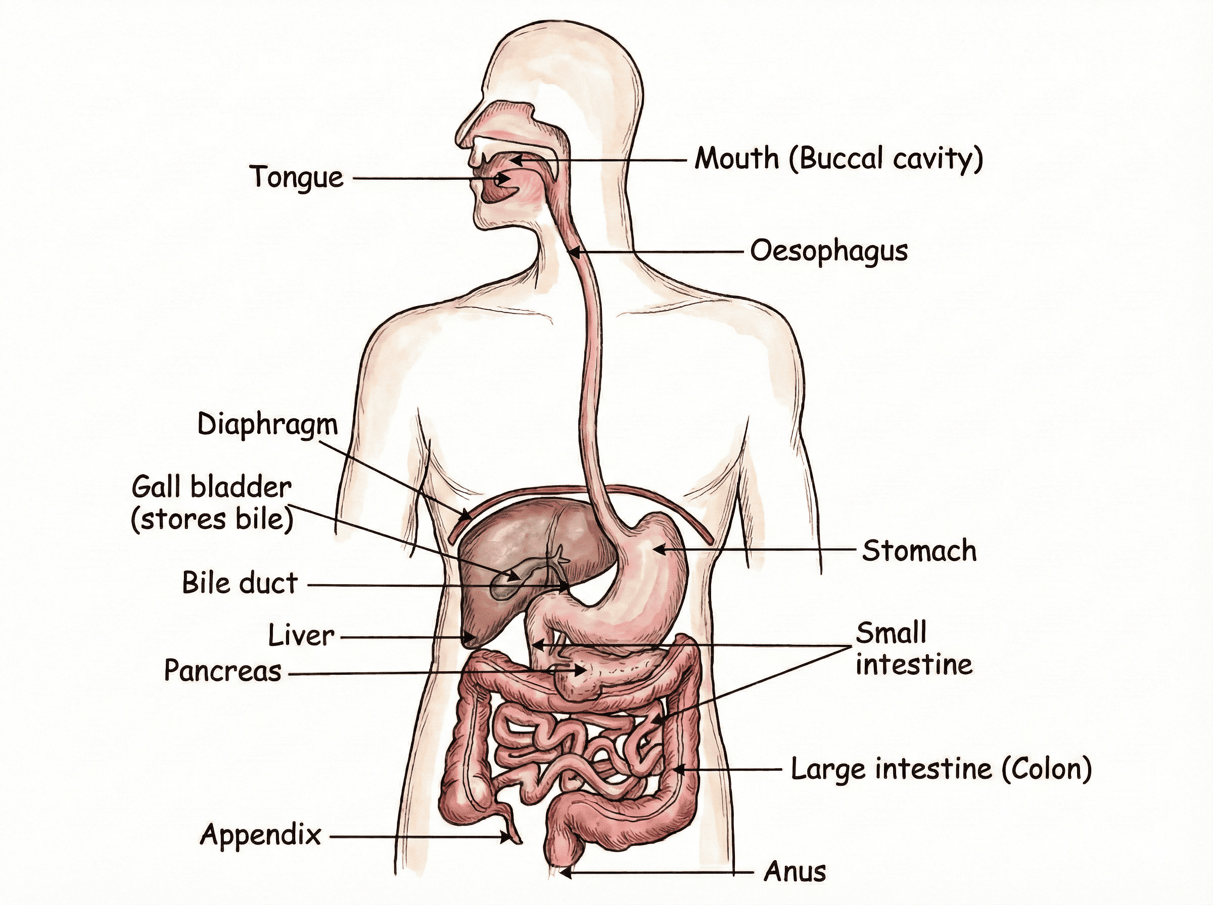
Figure 22.5: Anatomy of the Human Alimentary Canal and Associated Glands
Master Table: Human Digestive Enzymes & Secretions
| Organ / Gland |
Secretion |
Active Enzyme / Agent |
Biochemical Action |
| Mouth (Salivary Glands) |
Saliva ($\text{pH} \approx 6.8$) |
Salivary Amylase (Ptyalin) |
Breaks complex Starch into soluble disaccharide Maltose. |
| Stomach (Gastric Glands) |
Gastric Juice |
Hydrochloric Acid ($\mathrm{HCl}$) |
Creates acidic medium ($\text{pH} \approx 1.5 - 2.5$), activates pepsinogen to pepsin, and kills ingested pathogens. |
| Pepsin |
Digests Proteins into smaller peptones and proteoses. |
| Mucus |
Coats and shields the inner mucosal wall from excoriation by corrosive $\mathrm{HCl}$. |
| Liver |
Bile Juice (No enzymes, alkaline) |
Bile Salts & Pigments |
Emulsification of Fats: Breaks large fat globules into minute droplets; neutralizes acidic chyme to alkaline ($\text{pH} \approx 7.8 - 8.4$). |
| Pancreas |
Pancreatic Juice |
Pancreatic Amylase |
Hydrolyzes remaining starch into maltose. |
| Trypsin |
Digests peptones and proteins into peptides (active in alkaline medium). |
| Pancreatic Lipase |
Hydrolyzes emulsified lipids/fats into fatty acids and glycerol. |
| Small Intestine (Crypts) |
Intestinal Juice (Succus Entericus) |
Peptidases, Maltase, Sucrase, Intestinal Lipase |
Final digestion: Peptides $\rightarrow$ Amino Acids; Disaccharides $\rightarrow$ Glucose; Fats $\rightarrow$ Fatty Acids + Glycerol. |
Specialized Adaptations of the Small Intestine for Absorption:
The small intestine is the principal site of absorption of digested nutrients. It exhibits distinct structural marvels:
- Intestinal Villi: The inner mucosal lining contains millions of microscopic, finger-like projections called villi, which multiply the effective absorptive surface area exponentially.
- Rich Capillary Network: Each villus contains a dense network of blood capillaries that absorb water-soluble monosaccharides (glucose) and amino acids directly into the bloodstream.
- Lacteals: The center of each villus is occupied by a central lymphatic capillary termed a lacteal, which absorbs insoluble digested fatty acids and glycerol into the lymphatic circulation.
Large Intestine & Egestion
The undigested food mass passes into the Large Intestine (colon), whose walls absorb excess water and residual salts. The remaining semi-solid waste is compacted and stored temporarily in the rectum until it is expelled through the anus, an exit regulated precisely by the anal sphincter.
3. Respiration: Energy Release & Gas Exchange
Respiration is the cellular biochemical catabolism of organic nutrients to liberate free energy, which is trapped in high-energy phosphate bonds of ATP (Adenosine Triphosphate)—the universal energy currency of cells.
Crucial Distinction: Breathing vs. Cellular Respiration
Breathing (External Respiration): A purely mechanical, physical process involving the muscular inhalation of fresh air rich in oxygen and exhalation of air enriched with carbon dioxide. No energy is released, and no enzymes are involved.
Cellular Respiration (Internal Respiration): A multi-step enzymatic, biochemical process taking place within cells where glucose molecules are broken down to yield metabolic energy in the form of ATP.
A. Pathways of Glucose Breakdown
The initial phase of glucose breakdown—Glycolysis—occurs universally in the cytoplasm of all living cells without requiring molecular oxygen. During glycolysis, one molecule of 6-carbon glucose is cleaved into two molecules of 3-carbon Pyruvate, producing a net yield of 2 ATP.
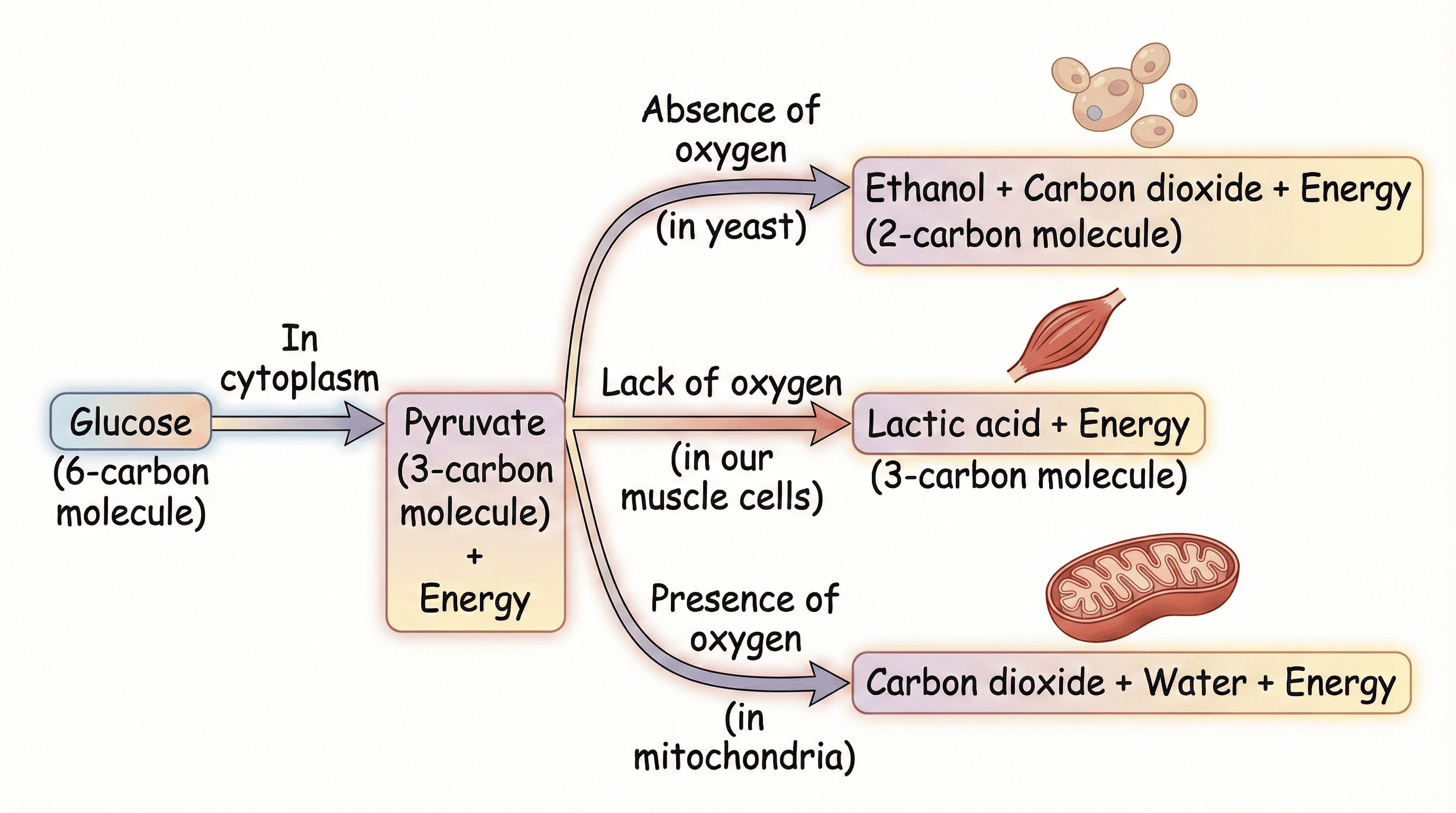
Figure 22.6: The Three Metabolic Fates of Pyruvate in Living Organisms
| Pathway |
Conditions & Site |
End Products |
Energy Harvest |
| Fermentation (Anaerobic in Yeast) |
Absence of $\mathrm{O_2}$, occurs entirely in Cytoplasm. |
Ethanol ($\mathrm{C_2H_5OH}$) + Carbon Dioxide ($\mathrm{CO_2}$) |
Low (2 ATP per glucose) |
| Anaerobic in Human Muscle |
Lack of $\mathrm{O_2}$ during sudden, heavy physical exercise (Cytoplasm). |
Lactic Acid ($\mathrm{C_3H_6O_3}$) |
Low (2 ATP per glucose)
Causes muscle cramps! |
| Aerobic Respiration |
Presence of abundant $\mathrm{O_2}$, occurs in Mitochondria. |
Carbon Dioxide ($\mathrm{CO_2}$) + Water ($\mathrm{H_2O}$) |
High (36 - 38 ATP per glucose) |
During strenuous physical sprinting or intense muscular exertion, the demand for oxygen outpaces the delivery capacity of the circulatory system. Muscle cells switch transiently to anaerobic respiration to produce emergency ATP. The resulting accumulation of lactic acid in muscle fibers causes localized pain, fatigue, and muscular cramps. Taking a hot water bath or gentle massage restores blood circulation, accelerating oxygen supply to oxidize lactic acid back into $\mathrm{CO_2}$ and water.
B. Human Respiratory System
Terrestrial human respiration involves a continuous branching network of air conducting passages leading into deeply protected internal lungs.
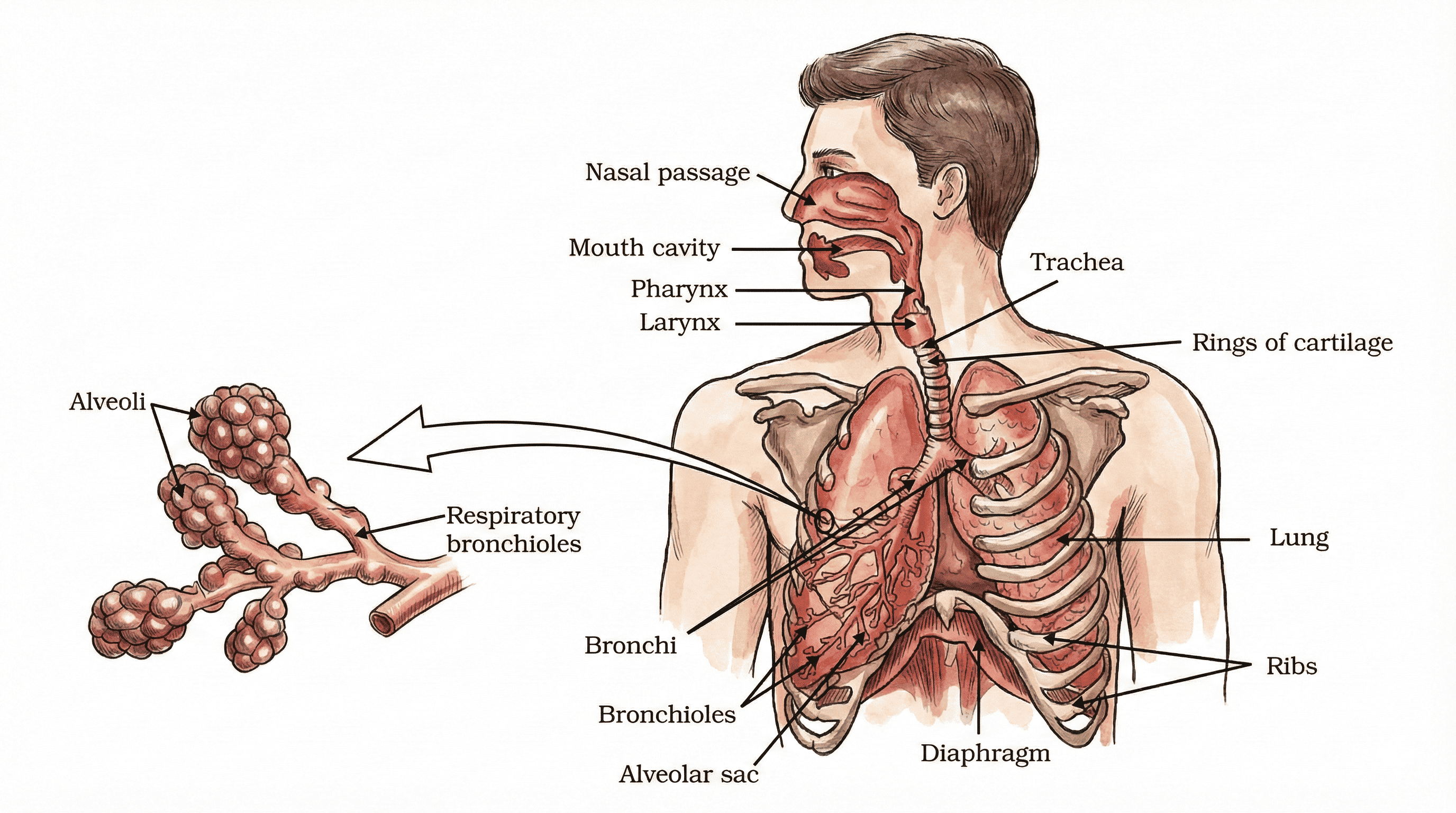
Figure 22.7: Detailed Structural Anatomy of the Human Respiratory Tract and Alveoli
Structural Journey of Air Inhalation:
- Nostrils: Inhaled air enters through the external nares, where fine hair and a viscous mucous lining filter out airborne dust particles, microbes, and pollutants while warming and humidifying the airstream.
- Pharynx & Larynx: Common passage for air and food. The epiglottis acts as a cartilaginous lid over the glottis to prevent food from entering the trachea during swallowing. The larynx houses vocal cords for phonation.
- Trachea (Windpipe): A rigid tube supported by C-shaped cartilaginous rings that prevent the airway from collapsing when negative pressure develops during inspiration.
- Bronchi & Bronchioles: Trachea bifurcates into right and left primary bronchi, which branch repeatedly into primary, secondary, tertiary bronchi and microscopic terminal bronchioles.
- Alveolar Sacs (Alveoli): Millions of microscopic, cup-shaped balloon-like sacs at the terminus of bronchioles. If flattened, human alveoli would cover a staggering surface area of nearly $80\,\text{m}^2$ (equivalent to a tennis court!). Their ultra-thin, single-cell thick squamous walls are wrapped in an extensive capillary mesh, creating an ideal interface for rapid gas diffusion.
- Inhalation (Inspiration): The muscular dome-shaped diaphragm contracts and flattens downwards, while the external intercostal muscles contract, lifting the rib cage upwards and outwards. The thoracic volume increases, causing intrapulmonary air pressure to drop below atmospheric pressure. Outside air rushes in to inflate the lungs.
- Exhalation (Expiration): The diaphragm relaxes and arches upward into its dome shape; intercostal muscles relax, allowing ribs to fall downward and inward. Thoracic volume contracts, raising internal pressure and pushing stale air out.
- Residual Volume: During normal breathing cycles, the lungs always retain a mandatory volume of residual air (approx. $1100 - 1200\,\text{mL}$) to allow continuous gas exchange even between successive breaths.
The Respiratory Pigment: Hemoglobin
In large multicellular animals like humans, passive diffusion is vastly insufficient—calculations show it would take nearly 3 years for an oxygen molecule to diffuse from our lungs to our toes! Nature overcomes this via the iron-rich respiratory pigment Hemoglobin ($\mathrm{Hb}$) packed inside Red Blood Cells (RBCs):
- $\mathrm{Hb}$ exhibits high chemical affinity for $\mathrm{O_2}$, binding reversibly to form Oxyhemoglobin ($\mathrm{Hb(O_2)_4}$).
- Carbon dioxide is significantly more water-soluble than oxygen, so it is transported primarily dissolved in the liquid blood plasma as bicarbonate ions ($\mathrm{HCO_3^-}$).
Investigation 3: Demonstration of CO₂ Exhalation
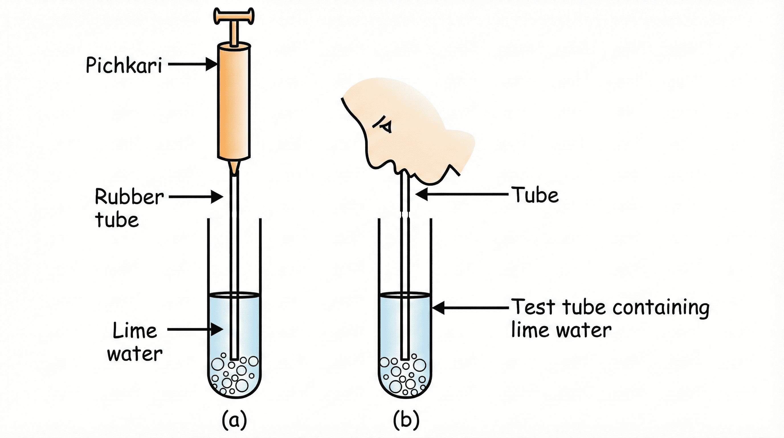
Figure 22.8: Comparing lime water milky reaction with exhaled air vs ambient pichkari air
Procedure: Take two test tubes containing freshly prepared clear Lime Water [$\mathrm{Ca(OH)_2}$]. In Test Tube A, pump ambient atmospheric air using a syringe/pichkari. In Test Tube B, blow exhaled breath gently through a straw.
Observation: Lime water in Test Tube B turns milky almost instantly due to insoluble Calcium Carbonate precipitation:
$$\mathrm{Ca(OH)_2} + \mathrm{CO_2} \rightarrow \mathrm{CaCO_3}\downarrow \text{(Milky)} + \mathrm{H_2O}$$
Test Tube A requires prolonged pumping to show faint cloudiness.
Inference: Expired breath contains a markedly higher concentration of carbon dioxide than atmospheric air, verifying cellular metabolic waste release.
4. Transportation in Animals and Plants
Multicellular organisms require efficient, high-speed internal transit networks to supply nutrients, hormones, and oxygen to all cells and remove metabolic wastes.
A. Transportation in Humans (Circulatory System)
The human circulatory system comprises three coordinated components: the circulating fluid (Blood & Lymph), the pumping organ (Heart), and the delivery conduit network (Blood Vessels).
Components of Human Blood:
- Plasma (55%): Straw-colored viscous fluid containing $90-92\%$ water, mineral ions, albumin, globulins, fibrinogen, hormones, digested food nutrients, and nitrogenous wastes.
- Red Blood Cells (Erythrocytes): Biconcave, enucleated disc cells packed with Hemoglobin to transport oxygen.
- White Blood Cells (Leukocytes): Nucleated amoeboid defense cells that synthesize antibodies and engulf foreign microbial pathogens (phagocytosis).
- Platelets (Thrombocytes): Cell fragments critical for blood coagulation at injury sites, preventing fatal hemorrhage and loss of vascular pressure.
Anatomy and Internal Pumping of the Human Heart
The human heart is a hollow, conical, muscular organ roughly the size of a clenched fist (approx. $300\,\text{g}$), enclosed in a protective double-walled sac called the pericardium.
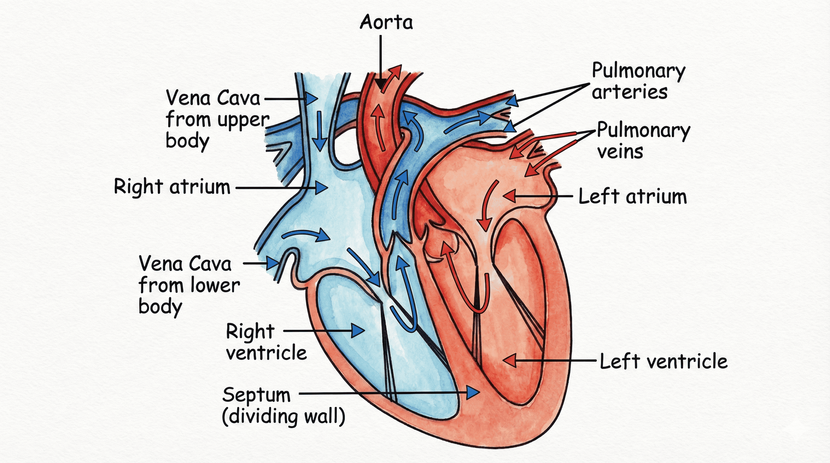
Figure 22.9: Internal Anatomy and Four-Chambered Architecture of the Human Heart
- Right Atrium & Right Ventricle: Handle deoxygenated blood arriving from systemic body tissues.
- Left Atrium & Left Ventricle: Handle freshly oxygenated blood arriving from pulmonary capillaries.
- Interventricular & Interatrial Septum: A thick, impervious muscular wall completely separating the left and right halves, preventing any mixing of oxygenated and deoxygenated blood. This separation ensures high physiological efficiency required by warm-blooded endotherms (birds and mammals) to maintain a constant internal body temperature.
- Thicker Ventricular Walls: Ventricles have substantially thicker, highly muscular walls than atria because they must pump blood under strong hydrostatic pressure across long distances (to the lungs and entire body), whereas atria merely push blood into adjacent ventricles.
- One-Way Valves: The Tricuspid Valve (right side) and Bicuspid/Mitral Valve (left side) prevent backflow of blood into atria during ventricular contraction (systole). Semilunar valves safeguard the exits into the aorta and pulmonary artery.
Double Circulation in Humans
Humans exhibit Double Circulation, meaning blood traverses through the heart twice during each complete circuit of the body:
.png)
Figure 22.10: Complete Circuit of Double Circulation: Pulmonary and Systemic Pathways
- Pulmonary Circuit: Deoxygenated blood from the body enters the Right Atrium via the Vena Cava $\rightarrow$ passes into Right Ventricle $\rightarrow$ pumped via Pulmonary Artery to the Lungs $\rightarrow$ oxygenated in alveolar capillaries $\rightarrow$ returned via Pulmonary Veins into the Left Atrium.
- Systemic Circuit: Oxygenated blood from Left Atrium passes into Left Ventricle $\rightarrow$ pumped forcefully via the massive Aorta to all systemic organs and tissues $\rightarrow$ deoxygenated blood is recollected into the superior and inferior Vena Cava $\rightarrow$ drains into Right Atrium.
Comparative Matrix: Arteries vs. Veins vs. Capillaries
| Feature |
Arteries |
Veins |
Capillaries |
| Direction of Flow |
Carry blood away from heart to organs. |
Carry blood towards the heart from organs. |
Connect arterioles with venules. |
| Nature of Blood |
Oxygenated (Exception: Pulmonary Artery). |
Deoxygenated (Exception: Pulmonary Vein). |
Gradual transition from oxygenated to deoxygenated. |
| Vessel Wall |
Thick, muscular, highly elastic. |
Thin, less muscular, less elastic. |
Extremely thin (single layer of endothelium). |
| Internal Valves |
Absent (blood flows under high pulsatile pressure). |
Present (unidirectional pocket valves prevent backward flow). |
Absent. |
| Lumen Diameter |
Narrow lumen. |
Wide lumen. |
Microscopic (allows RBCs to pass in single file). |
Lymphatic System (Tissue Fluid)
At the capillary beds, some amount of blood plasma, proteins, and leukocytes leak through intercellular endothelial pores into surrounding tissue spaces, forming Tissue Fluid or Lymph. Lymph is colorless and contains less protein than blood plasma. It enters microscopic lymphatic capillaries, which unite into larger lymph vessels that eventually empty into large veins near the neck. Lymph returns extravasated fluid to the bloodstream and transports absorbed dietary fats from intestinal lacteals.
B. Transportation in Plants
Plants are stationary and have low metabolic rates compared to animals, so they do not require rapid muscular pumps. Transport is carried out through two distinct specialized vascular complexes:
| Feature |
Xylem |
Phloem |
| Substances Transported |
Water and dissolved mineral salts (inorganic ions). |
Soluble organic products of photosynthesis (chiefly Sucrose) and amino acids. |
| Direction of Flow |
Unidirectional (strictly upward from roots to stems and leaves). |
Bidirectional (from source leaves to storage sinks and developing buds). |
| Conducting Elements |
Tracheids and Xylem Vessels (dead, hollow, lignified tubes at maturity). |
Sieve Tubes and Companion Cells (living cells with perforated sieve plates). |
| Energy Expenditure |
Driven by physical forces (Transpiration Pull and Root Pressure); No ATP used. |
Active transport; requires chemical energy in the form of ATP. |
- Root Pressure (Night-time Driver): Root cortical cells actively pump mineral ions into root xylem. This creates an osmotic concentration difference between the root and soil, drawing water into root xylem and generating a positive hydrostatic root pressure. This force is sufficient to move water upward in short herbaceous plants.
- Transpiration Pull (Daytime Driver): During daytime, continuous evaporation of water molecules through open leaf stomata creates an intense negative suction force (transpiration pull). Because water exhibits strong cohesive forces (water-to-water attraction) and adhesive forces (water-to-xylem wall attraction), an unbroken capillary water column is pulled up to the highest canopies of gigantic trees (like Sequoia, reaching over 100 meters!).
5. Excretion: Waste Elimination & Osmoregulation
Metabolic activities, particularly protein catabolism, generate hazardous nitrogenous wastes like ammonia, urea, and uric acid. Excretion is the biological process of removing these toxic metabolic wastes and balancing water/electrolyte levels (osmoregulation).
A. Human Excretory System
The human urinary system consists of a pair of kidneys, a pair of ureters, a urinary bladder, and a urethra.
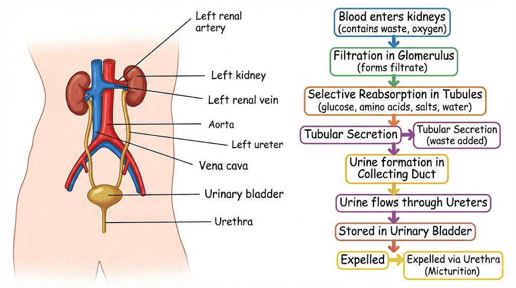
Figure 22.11: Gross Anatomical Layout of the Human Excretory System
Organs of Excretion:
- Kidneys: Pair of reddish-brown, bean-shaped organs located in the dorsal abdominal cavity on either side of the vertebral column.
- Ureters: Long, slender muscular tubes originating from the renal pelvis that convey urine from each kidney to the urinary bladder via peristaltic waves.
- Urinary Bladder: A distensible, muscular reservoir lined with transitional epithelium that holds urine under voluntary nervous control until micturition.
- Urethra: The terminal muscular canal discharging urine outside the body, guarded by internal (involuntary) and external (voluntary) urethral sphincters.
Structure and Functional Mechanism of the Nephron
The structural and functional filtration unit of the kidney is the Nephron. Each human kidney contains approximately 1 to 1.2 million microscopic nephrons.
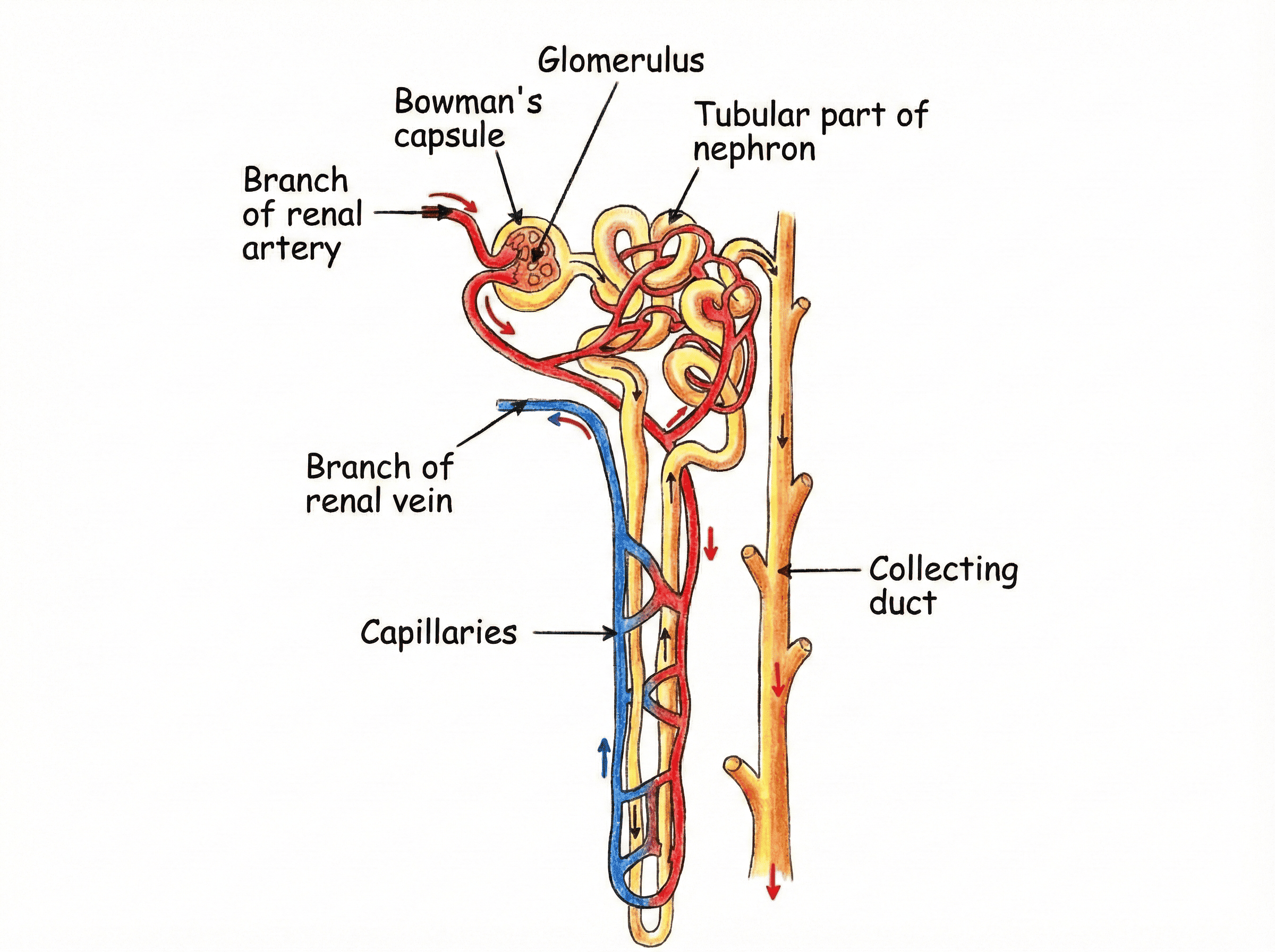
Figure 22.12: Microscopic Structural Organization of a Nephron and Peritubular Capillaries
The Three Sequential Stages of Urine Formation
- 1. Glomerular Ultrafiltration: Blood enters the Glomerulus (a dense capillary knot) via the wide Afferent Arteriole under high hydrostatic pressure and exits through the narrow Efferent Arteriole. This pressure differential forces water, glucose, amino acids, urea, and electrolytes through the podocyte filtration slits into Bowman's Capsule. Blood cells and large plasma proteins cannot pass and remain in the bloodstream.
The initial glomerular filtrate generated in both kidneys equals a massive 180 Litres per day!
- 2. Selective Reabsorption: As the filtrate travels down the Proximal Convoluted Tubule (PCT), Loop of Henle, and Distal Convoluted Tubule (DCT), all useful substances—including $100\%$ of glucose, amino acids, and major mineral salts along with most of the water—are selectively reabsorbed back into the surrounding peritubular capillaries.
Because of intensive selective reabsorption, out of 180 L of initial filtrate, only 1.5 to 2 Litres of concentrated urine is excreted daily!
- 3. Tubular Secretion: Cells of the renal tubules selectively secrete surplus hydrogen ions ($\mathrm{H^+}$), potassium ions ($\mathrm{K^+}$), and ammonium ions from peritubular capillary blood into the tubular lumen to maintain precise blood pH and electrolyte equilibrium.
Hemodialysis: The Artificial Kidney
In cases of acute renal failure due to injury, diabetes, or hypertension, toxic urea accumulates in the blood (uremia), which is fatal if untreated. Hemodialysis is a medical procedure using an artificial dialyzer unit:
- Blood pumped from a patient's artery is cooled, mixed with anticoagulant (heparin), and passed through a coiled semipermeable cellophane tubing suspended in a dialyzing fluid.
- The dialyzing solution has identical osmotic and electrolyte composition to normal blood plasma, but contains zero nitrogenous waste.
- Toxic wastes (urea, uric acid) rapidly diffuse across the cellophane membrane down their concentration gradient into the dialyzing fluid.
- The purified blood is warmed, treated with anti-heparin, and safely infused back into the patient's vein.
- Crucial Difference: The artificial kidney performs ultrafiltration and diffusion, but performs no selective reabsorption like a natural nephron.
B. Excretion in Plants
Plants possess completely different excretory strategies compared to animals because their metabolic rates are much slower and they generate negligible amounts of toxic nitrogenous waste:
- Gaseous Wastes: Excess $\mathrm{O_2}$ generated during photosynthesis and $\mathrm{CO_2}$ from cellular respiration diffuse directly out into the atmosphere through stomata in leaves and lenticels in woody stems.
- Excess Water: Removed continuously as water vapor via transpiration.
- Storage in Aging Tissues: Many plant wastes are deposited as harmless crystals (like calcium oxalate raphides) within cellular vacuoles, or stored in dead heartwood, shedding bark, and aging leaves that eventually fall off.
- Gums and Resins: Harmful secondary metabolic byproducts are sequestered safely as insoluble gums and resins in old, non-functional xylem vessels.
- Root Exudates: Some metabolic wastes are actively secreted directly into the rhizosphere soil through root epidermis.
6. High-Yield Examination Summary
Quick Revision Points for Board Exams
- Photosynthesis Site: Chloroplast (Thylakoids for light reaction; Stroma for dark reaction).
- Source of Oxygen: Photolysis of water ($H_2O$), not from carbon dioxide.
- Emulsification: Mechanical breakdown of large fat droplets into tiny micelles by bile salts to increase enzyme surface area.
- Protein Digestion: Starts in stomach with Pepsin (acidic $\text{pH}$); completed in small intestine with Trypsin and peptidases (alkaline $\text{pH}$).
- Site of Complete Digestion: Small Intestine (specifically duodenum and jejunum).
- Cramps Cause: Lactic acid accumulation during anaerobic respiration in muscle cells due to oxygen deficit.
- Pumping Pressures: Normal human blood pressure is 120/80 mm Hg (Systolic/Diastolic), measured using a Sphygmomanometer.
- Double Circulation Necessity: Ensures complete separation of oxygenated and deoxygenated blood, providing peak metabolic efficiency for homeothermy (warm-bloodedness).
- Translocation: Movement of sucrose via phloem driven actively by osmotic water pressure using ATP.
- Nephron Filtration Unit: Malpighian body (Glomerulus + Bowman's Capsule).
7. Concept Review & Board Exam Practice
Question 1: Analytical Reasoning (3 Marks)
Q: Why is the rate of breathing in aquatic organisms much faster than that seen in terrestrial organisms?
Model Answer: Terrestrial organisms breathe atmospheric air, which contains a high concentration of oxygen (approximately $21\%$). In contrast, aquatic organisms utilize oxygen dissolved in water. The solubility of oxygen in water is very low compared to air. Consequently, to acquire the requisite amount of oxygen needed for cellular metabolism, aquatic animals (like fishes) must pump water across their gills much more rapidly, resulting in a substantially higher breathing rate.
Question 2: Physiological Comparison (3 Marks)
Q: Differentiate between the functions of Xylem and Phloem in plants with respect to substances transported, mechanism, and energy requirement.
Model Answer:
- Xylem: Transports water and inorganic minerals unidirectionally from roots upward. Transport is driven entirely by physical gradients (transpiration pull and root pressure) and does not consume metabolic cellular energy (ATP).
- Phloem: Transports synthesized food products (sucrose) and amino acids bidirectionally from source leaves to storage or growing tissues (translocation). Solutes are actively loaded into sieve tubes using cellular energy from ATP, which drives mass flow via osmotic pressure gradients.
Question 3: Comprehensive Mechanism (5 Marks)
Q: Describe the structural organization of a Nephron and explain how urine is formed in the human kidney. Why is the volume of excreted urine much less than the initial filtrate formed?
Model Answer:
- Structural Organization: A nephron consists of a cup-shaped Bowman's Capsule enclosing a capillary cluster called the Glomerulus, followed by a coiled Proximal Convoluted Tubule (PCT), a hairpin-shaped Henle's Loop, and a Distal Convoluted Tubule (DCT) opening into a common Collecting Duct.
- Stages of Urine Formation:
- Glomerular Ultrafiltration: High hydrostatic pressure forces water, glucose, salts, and urea through glomerulus into Bowman's capsule, creating initial filtrate ($\approx 180\,\text{L/day}$).
- Selective Reabsorption: As filtrate flows along PCT and Henle's loop, essential substances (all glucose, amino acids, essential salts, and $99\%$ of water) are reabsorbed back into peritubular capillaries.
- Tubular Secretion: Surplus ions ($\mathrm{H^+}$, $\mathrm{K^+}$, ammonia) are secreted from blood into filtrate to regulate blood pH and electrolyte balance.
- Explanation of Volume Difference: Although $180\,\text{L}$ of filtrate is filtered daily, about $178 - 178.5\,\text{L}$ ($99\%$) is selectively reabsorbed by the tubular nephron walls back into the bloodstream. Therefore, only $1.5 - 2\,\text{L}$ of concentrated urine is excreted daily.

 VARDAAN COMET