
ICSE Class 10 Biology • Chapter 11 • Detailed Master Notes
Reproduction is the key point of the continuance of life on earth. In a way, all other systems and processes in the body are for supporting the reproduction process.
Reproduction is the formation of new individuals by sexual or asexual means, which can repeat the process in their own turn.
(i) Asexual reproduction: The reproductive units may under asexual type of reproduction consist of any portion of the parent body. The size of the units may range from the whole to a small fragment of the parent body.
(ii) Sexual reproduction: The gametes (sex cells - egg and sperm) are very small (microscopic in human beings). The gametes must normally unite (fertilisation) to produce a zygote which, through a process of development, finally becomes the offspring.
| Asexual reproduction | Sexual reproduction |
|---|---|
| 1. No mixing of genetic material, therefore, no or less variation in offsprings. | 1. Genetic mixing, increased variation. |
| 2. No gametes are formed. | 2. Gametes are formed. |
| 3. Normally more offspring. | 3. Fewer offspring. |
| 4. Only one parent is involved. | 4. Usually, two parents (male and female) are involved. |
| 5. It is a rapid process during favourable conditions. | 5. Slower process. |
The organs of the reproductive system are divided into primary and accessory parts:
The male reproductive system consists of the following organs:
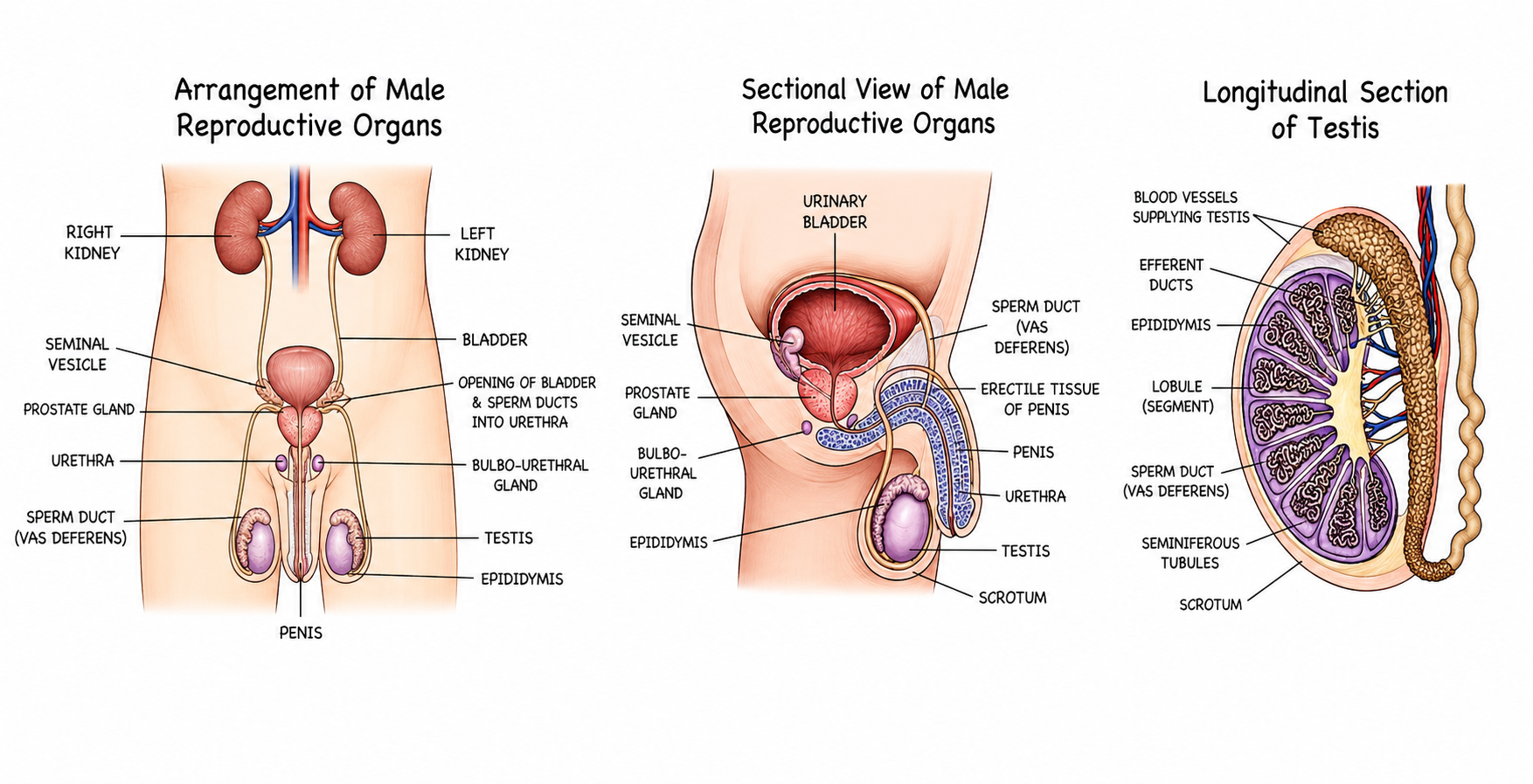
A. Location of testes:
Temperature regulation in the testes: Sperms are produced in the testes at a temperature 2 to 3°C lower than that of the body. This temperature is regulated through movements of the scrotum wall. When it is too hot, the skin loosens so testes hang away; when cold, skin contracts to draw them closer for warmth.
B. Structure of testis:
Each testis is internally partitioned into 15-20 lobules. Each lobule contains:
The sperm duct (vas deferens) from each testis travels upward into the abdomen passing through an inguinal canal.
The mature sperms pass into a network of tubes (efferent ducts) which join a small tubular knot, the epididymis fitting like a cap on the upper pole of the testis. Epididymis stores sperms for some days during which they mature and become motile.
The penis lies in front of the scrotum, cylindrical in shape, serves for the passing out of both semen and urine. It has highly vascular erectile tissues.
The female reproductive system consists of the following reproductive organs:
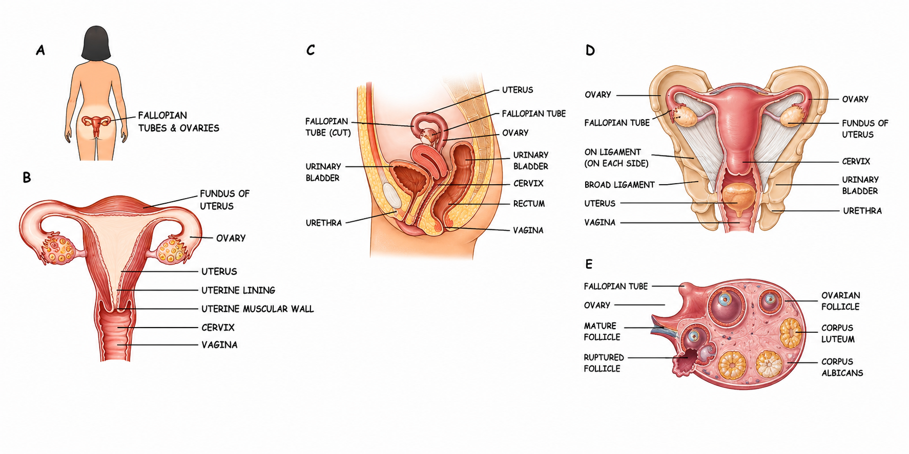
The two ovaries are small ovoid bodies. Their peripheral part produces ova or the eggs. Normally, only one egg matures in each ovary every alternate month. A maturing egg contained in a cellular sac is called the follicle. As the egg grows larger, the follicle enlarges, gets filled with fluid, and is called the Graafian follicle. Oogenesis is the process in which the ova-producing cells give rise to the mature ovum.
Ovulation: It is the rupture of the follicle releasing the egg. The released egg is picked up by fimbriae (cilia) of the oviducal funnel.
The remnant of the follicle persists to convert into a yellow mass called corpus luteum. It secretes two hormones:
The two oviducts, also called Fallopian tubes or uterine tubes, are about 12 cm long. Each has a funnel-shaped opening called the oviducal funnel. Cilia lining the funnel help push the ovum into the oviduct. Muscular contractions (peristalsis) push the egg down into the uterus.
The uterus is a hollow pear-shaped muscular organ situated in the pelvic cavity between the urinary bladder and rectum. It has an upper wider portion and a small lower constricted part, the cervix or neck.
The vagina is a muscular tube (10-15 cm long) starting from the lower end of the uterus up to the outside. It receives the male penis during copulation and allows passage of the baby during childbirth.
The reproductive period of the human female continues from about the age of 13 years to 45-50 years. This period is marked by a characteristic event repeated almost every month (28 days with minor variation) in the form of a menstrual flow.
Menarche: The onset of menstruation in a young female at about the age of 13 years (arche: beginning).
Menopause: The permanent stoppage of menstruation at about the age of 45 years (pause: stop).
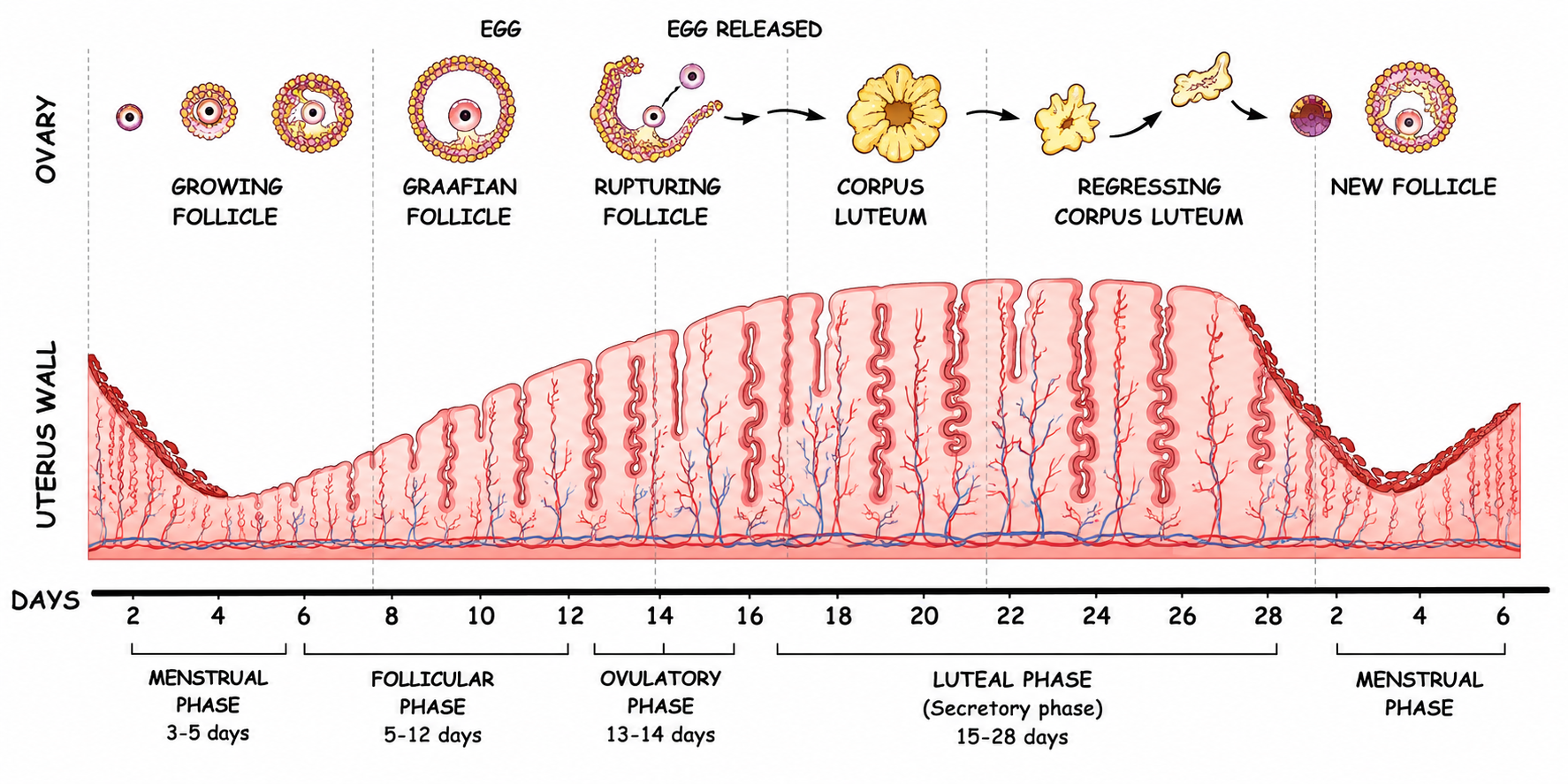
What if fertilisation does not occur?
If there is no fertilisation, the egg disintegrates and the uterine lining restarts shedding on the 28th day. If the egg is fertilised, it gets implanted in the uterus wall and there is no menstrual discharge (pregnancy).
During copulation (mating or coitus), sperms are released in the vagina near the cervix. A single ejaculation contains about 400 million sperms. They actively swim through the cervix into the uterus and climb up to the oviducts. If there happens to be an ovum, it gets fertilised by just one sperm.
Fertilisation: The fusion of the male gamete (sperm) and female gamete (ovum) to form a zygote.
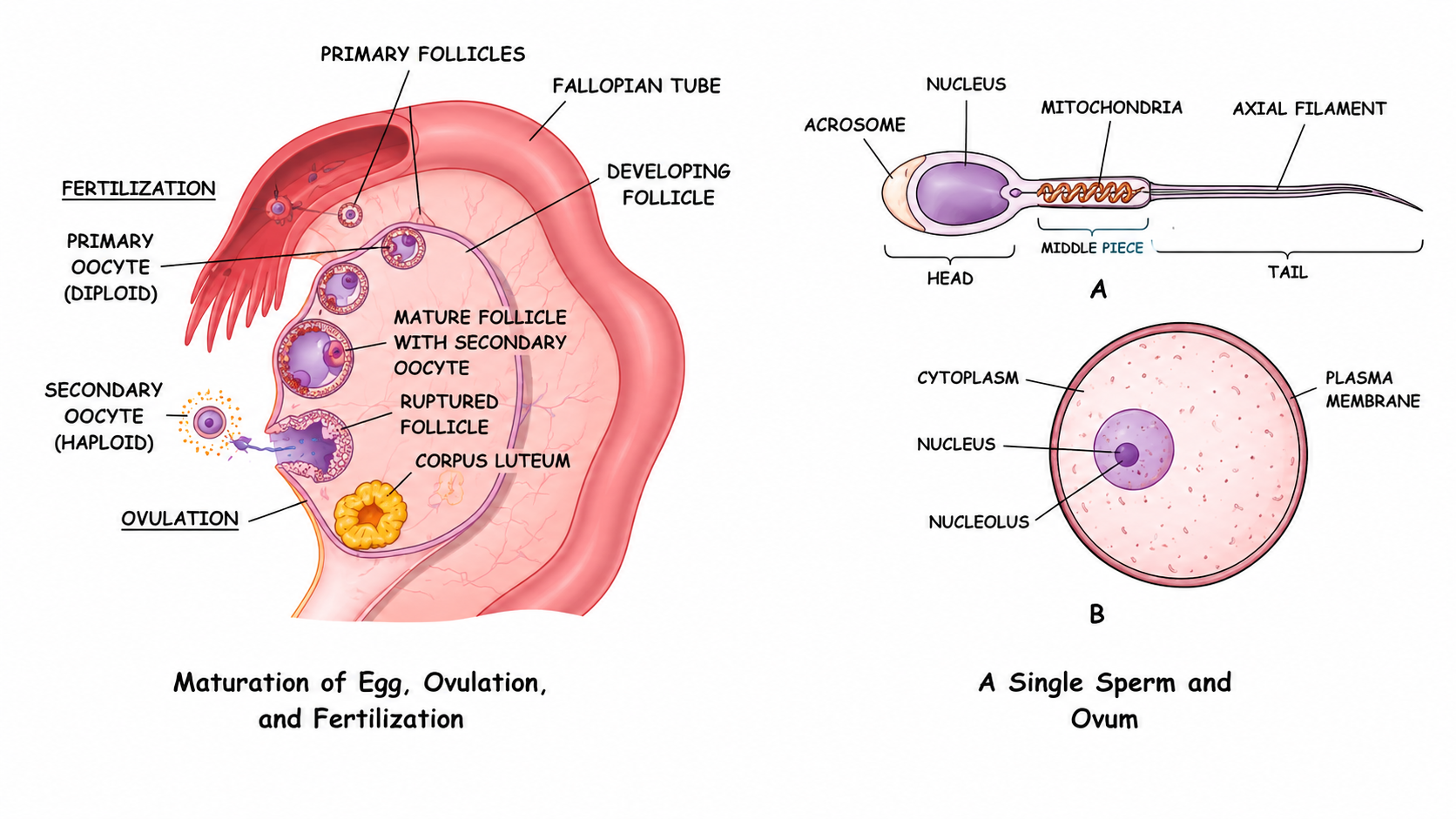
The fertilised egg (zygote) soon starts dividing (cleavage) into 2, 4, 8, 16 cells and so on. By the time it reaches the uterus, it has already formed a small hollow ball of numerous cells (blastocyst). This is a kind of embryo which forms a pit in the wall (endometrium) of the uterus and gets fixed in it in about a week's (5-7 days) time after ovulation.
Implantation: The process of fixation of the embryo (blastocyst) in the wall of the uterus. It produces the state of pregnancy.
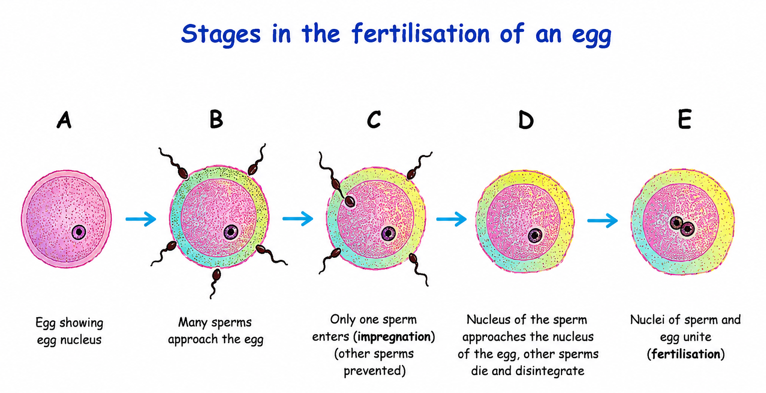
The sequence of development inside the oviduct and uterus:
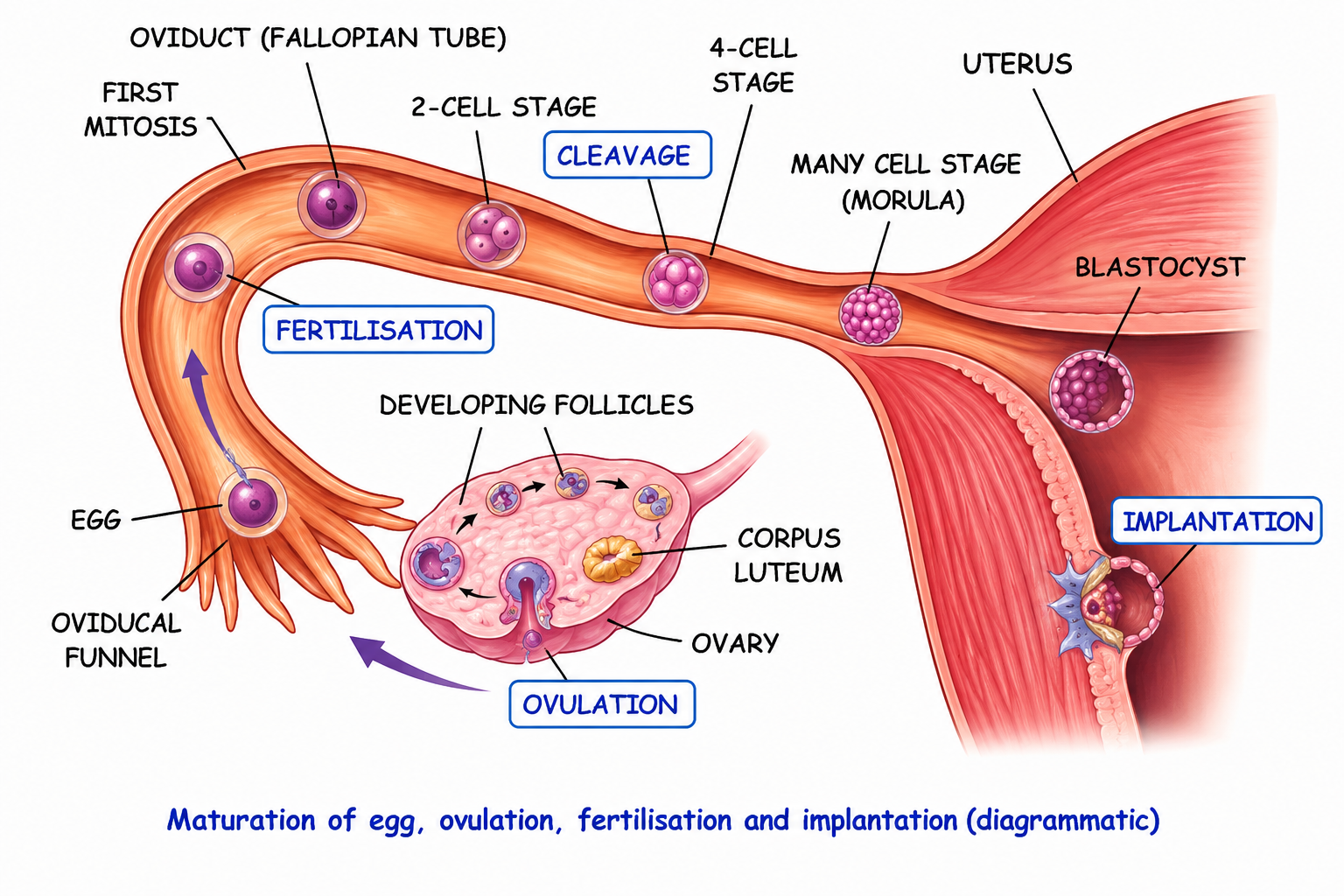
Amnion is a sac which develops around the embryo even before the formation of allantois. A fluid (amniotic fluid) fills the space between the amnion and the embryo.
The growing embryo or the foetus is a living organism. It needs food and oxygen. It excretes nitrogenous wastes and carbon dioxide which need to be continuously removed. These special functions are performed by a structure called placenta. Placenta is a disc-like structure attached to the uterine wall. A cord containing blood vessels connects the placenta with the foetus; this is called umbilical cord.
The placenta is formed of two sets of minute finger-like processes, the villi. One set of villi are given out by the uterine wall and the other set by an extension (allantois) from the embryo. The two sets of villi get interlocked but they never open into each other. Here, the blood of the embryo comes in close contact, but never mingles with the blood of the mother.
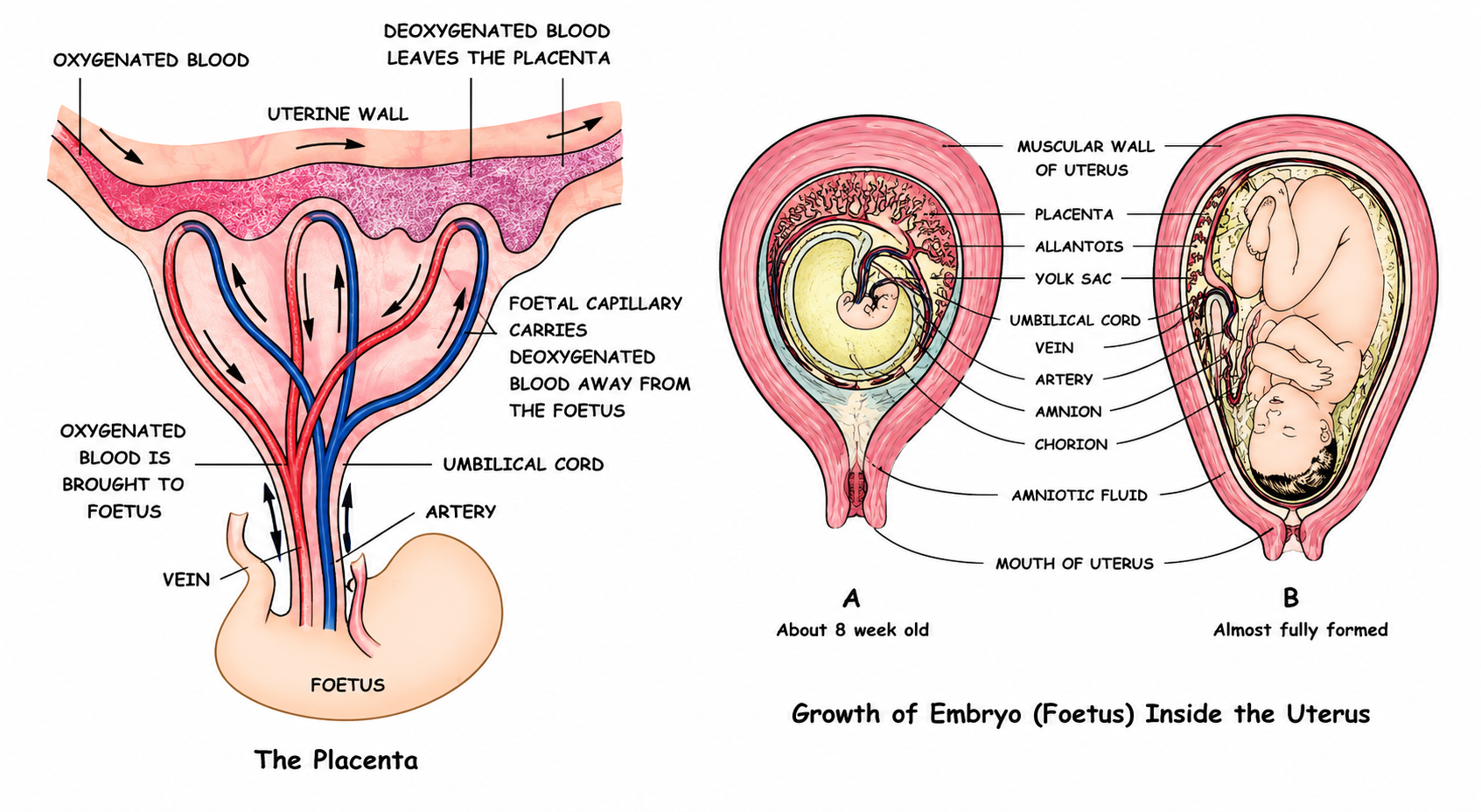
The placenta allows diffusion of substance from the mother to the foetus and from foetus to the mother as follows:
A. From mother to foetus (for utilisation):
B. From foetus to mother (for elimination):
The placenta is permeable to respiratory gases, nutrients and also antibodies but it does not allow the passage of germs from the mother to the foetus. Certain viruses like the HIV of AIDS can pass through the placenta if the mother is already infected. Secondly, if there was a direct continuity of blood between the mother and the foetus without a placenta, the relatively high pressure of the mother's blood would seriously damage the soft and delicate tissues of the foetus.
Placenta also acts as an endocrine gland: It produces the hormones called oestrogens and progesterone. Presence of progesterone in urine provides for certain tests of pregnancy.
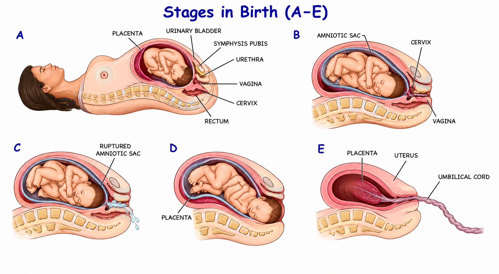
The full term of the development of an embryo in the uterus is called gestation. In humans, it lasts for about 280 days. At the time of birth, the baby is pushed out by the powerful contractions of the muscles of the uterus, head first.
In a few minutes, the blood vessels in the umbilical cord shrink and can be tied and cut. After about 15 minutes, the placenta breaks from the uterus and is expelled out as "after-birth." Uterus contracts and gradually comes back to normal in a few days.
The new-born "announces" its arrival by letting out sharp cry. This cry is the nature's clearing process of the baby's respiratory passage.
1. Fraternal Twins (Produced from two eggs)
Sometimes, two eggs are released from ovaries at a time and both may get fertilised to produce two individuals. Such twins produced from two eggs are called fraternal twins. They may be either both boys, or both girls, or one boy and one girl. They are biologically known as Diovular or Dizygotic.
2. Identical Twins (Produced from one egg)
Sometimes, a single fertilised egg may get split and separated into two parts during its early stages of cell division. Identical twins are either both boys or both girls; and for the same reason, they are very similar (identical) to each other. Yet, all is not similar in identical twins, for example fingerprints, birth marks and even handwritings are not similar.
Siamese Twins: These are conjoined twins produced from a single egg, who have failed to separate completely.