The Nervous System and Sense Organs
ICSE Class 10 Biology || Chapter 09 || Detailed
Master Notes
Every organism must become aware of what is going on around it and perform actions for its survival. Apart
from actions which provide adjustments to the external environment, there are many internal activities (like
breathing, digestion, heartbeat) of which we are unaware. All such actions have to be properly timed and
coordinated. This coordination occurs via two agencies: the Nervous System
and the Hormonal (Endocrine) System.
9.1 Need for a Nervous System
The nervous system performs several vital functions:
- Keeps us informed about the outside world through sense organs.
- Enables us to remember, think, and reason out.
- Controls and harmonises all voluntary muscular activities (e.g., running, writing).
- Regulates involuntary activities such as breathing, beating of the heart, and movements of the gut.
9.2 The Unit of the Nervous System: Neuron
Neuron (Nerve Cell): The structural and functional unit of the nervous system. The brain
consists of an estimated 100 billion neurons!
Structure of a Neuron
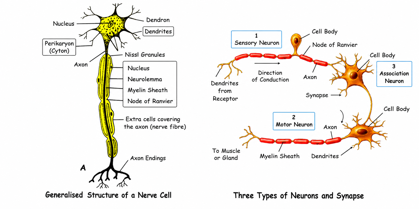
A typical neuron possesses three main parts:
- 1. Cyton (Cell Body / Perikaryon): The main body of the nerve cell containing a
prominent nucleus and cytoplasm. The cytoplasm contains characteristic darkly staining Nissl's granules (which help in protein synthesis).
- 2. Dendrites: Several short, branched extensions arising from the cyton. They
receive nerve impulses from other neurons and conduct them towards the cyton.
- 3. Axon: A single, long process extending from the cyton. It conducts impulses
away from the cyton to another neuron or an effector organ.
- Myelin Sheath: An insulating layer covering many axons, formed by
specialized Schwann cells. It speeds up the conduction of nerve impulses.
- Nodes of Ranvier: The gaps throughout the length of the myelin sheath where
the axon is exposed.
- Axon Endings (Terminals): The axon ends in fine branches bearing swollen
knob-like structures called Synaptic Knobs, which contain
neurotransmitters.
Some Basic Terms in Nervous Activity
- Stimulus: An agent or a sudden change of the external or internal environment that
results in a change in an organism or any of its body parts.
- Response: The change in an organism resulting due to stimulus.
- Impulse: A wave of irritability (electrical disturbance that sweeps over the nerve
cell).
- Receptors: The specialised epithelial cells which, on receiving the stimulus, set
up waves of impulses towards the central nervous system.
- Effectors: Muscles or glands which, on receiving the impulse from the brain or
spinal cord, contract or secrete substances.
Transmission of the Nerve Impulse
In the normal (resting) condition, the outer side of the nerve fibre carries positive (+) charge. This is
called the polarised state. This polarisation is due to more Na? ions outside the axon
membrane.
On stimulation (mechanical, electrical, chemical, or heat), the axon membrane at that spot becomes more
permeable to Na? ions which move inwards and cause loss of polarisation (depolarisation).
This is known as the excited region. The point of depolarisation becomes a stimulus for the
next neighbouring area of the membrane which in turn becomes depolarised.
Meanwhile, the previous area becomes repolarised due to active transport of Na? ions again
to the outside. This transport is achieved by what is called the "sodium pump" using energy
through ATP.
Conduction of nerve impulse is a wave of depolarisation followed by repolarisation.
Note: Electricity is conducted through a wire at a speed of about 150,000 km per second, but the
nerve impulse travels at a maximum speed of about 100 metres per second only.
Synapse
Synapse: The point of contact between the terminal branches of the axon of one neuron
and the dendrites of another neuron.
Nerve impulses travel across a synapse chemically through a neurotransmitter (e.g.,
Acetylcholine), ensuring the impulse travels in only one direction.
Types of Neurons
- Sensory Neurons: Carry impulses from sense organs (receptors) to the Central Nervous
System (Brain/Spinal Cord).
- Motor Neurons: Carry impulses from the CNS to the effector organs (muscles or glands)
causing them to react.
- Association (Interneurons): Located entirely within the CNS. They connect sensory and
motor neurons.
9.3 Nerves and Ganglia
A Nerve is a bundle of nerve fibres (axons) enclosed in a tubular medullary
sheath, acting like a communication cable. Nerves are of three types:
- Sensory Nerve: Contains only sensory fibres (e.g., Optic nerve from the eye to the
brain).
- Motor Nerve: Contains only motor fibres (e.g., nerve to the muscles of the eyeball).
- Mixed Nerve: Contains both sensory and motor fibres (e.g., Spinal nerves).
Ganglia: The aggregates of the cell bodies (cytons) from which the nerve fibres may
arise or enter into.
9.4 Divisions of the Nervous System
The nervous system has three main divisions:
- Central Nervous System (CNS): Consists of the Brain and Spinal Cord.
- Peripheral Nervous System (PNS): Consists of nerves emerging from the CNS (Cranial and
Spinal nerves).
- Autonomic Nervous System (ANS): Consists of a pair of chain of ganglia running along
the spinal cord (Sympathetic and Parasympathetic systems).
9.5 The Central Nervous System: Brain
The brain is the most complex organ, weighing about 1.35 kg (about 2% of body weight) but consuming 25% of
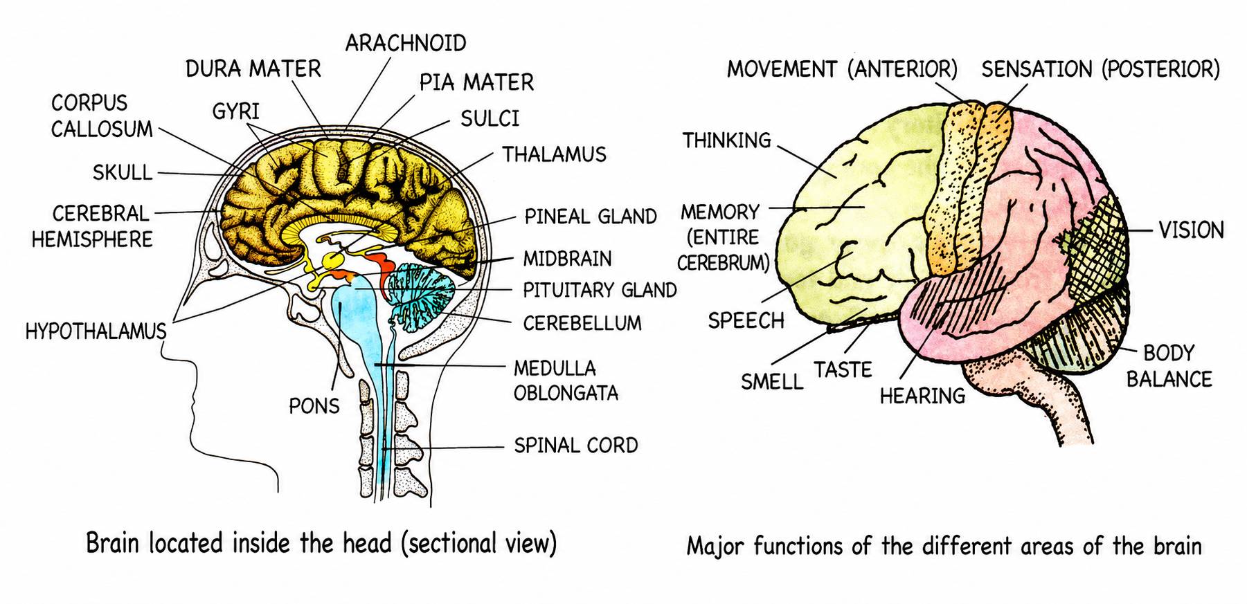
the total body oxygen. It is protected by the skull (cranium) and three membranous coverings called Meninges:
- Dura mater: Outermost, tough fibrous membrane.
- Arachnoid: Middle, delicate, web-like membrane.
- Pia mater: Innermost, highly vascular membrane closely investing the brain.
Cerebrospinal Fluid (CSF): The fluid filling the spaces between the meninges and the
ventricles of the brain. It acts as a shock absorber and provides nourishment.
Major Parts of the Brain
- Cerebrum: The largest part of the brain, divided into right and left
Cerebral Hemispheres.
- The two hemispheres are connected by a sheet of fibres called the Corpus Callosum.
- The outer portion (Cerebral Cortex) is Gray matter (cytons) and is highly
folded into convolutions (gyri) and grooves (sulci) to increase surface area to accommodate
more neurons.
- The inner portion is White matter (myelinated axons).
- Function: Seat of intelligence, consciousness, memory, willpower, and
controls all voluntary activities.
- Cerebellum ("Little Brain"): Located at the base under the cerebrum. It also has
outer gray and inner white matter.
- Function: Coordinates voluntary muscular movements and maintains body
balance/posture (e.g., if a drunk person staggers, the cerebellum is affected).
- Medulla Oblongata: The lowest part, continuing into the spinal cord.
- Function: Controls vital involuntary activities like heartbeat, breathing,
swallowing, coughing, and peristalsis. Injury here is usually fatal.
- Diencephalon: Contains the Thalamus (relay centre for pain and
pressure) and Hypothalamus (controls body temperature, water balance, hunger,
thirst, and regulates the pituitary gland).
9.6 The Central Nervous System: Spinal Cord
The spinal cord extends from the medulla oblongata down to the second lumbar vertebra. It is protected by the
vertebral column and the meninges.
Arrangement of Matter (Opposite to the Brain):
- Spinal Cord: White matter is on the OUTSIDE, Gray
matter is on the INSIDE (forming a butterfly or H-shape).
- Brain: Gray matter is on the outside (cortex), White matter is inside.
Functions of Spinal Cord: It is the center for reflexes below the neck, and it conducts
sensory impulses to the brain and motor impulses from the brain to effectors.
9.7 The Peripheral Nervous System (PNS)
The PNS connects the CNS to the rest of the body.
- Cranial Nerves: Emerge from the brain. Humans have 12
pairs (e.g., Optic nerve, Auditory nerve, Vagus nerve).
- Spinal Nerves: Emerge from the spinal cord. Humans have 31
pairs (8 cervical, 12 thoracic, 5 lumbar, 5 sacral, 1 coccygeal). All spinal nerves are mixed
nerves.
9.8 The Autonomic Nervous System (ANS)
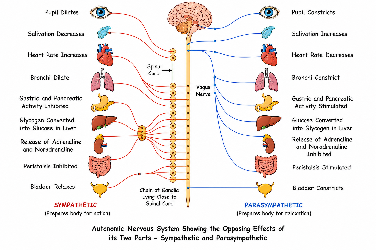
The ANS controls the involuntary activities of visceral organs (heart, blood vessels, glands). It consists of
two antagonistic systems:
- Sympathetic Nervous System: Prepares the body for violent action against abnormal
conditions (Fight or Flight). It is stimulated by adrenaline.
- Parasympathetic Nervous System: Restores normal conditions after the stress is over
(Rest and Digest).
Table 9.1: Effects of the two parts of autonomic nervous system
| Organs |
Sympathetic System |
Parasympathetic System |
| Heart |
Accelerates heart beat |
Retards heart beat |
| Blood vessels |
Constricts all blood vessels except coronary vessels which are dilated |
Dilates all blood vessels except coronary vessels which are constricted |
| Lungs |
Dilates bronchi and bronchioles |
Constricts bronchi and bronchioles |
| Intestines |
Peristalsis decreased |
Peristalsis increased |
| Urinary bladder |
Sphincter contraction, muscle relaxed |
Sphincter relaxation, muscle contraction (feeling to urinate) |
| Pupil of eye |
Dilation |
Constriction |
| Salivary glands |
Inhibits secretion of saliva (dryness of mouth) |
Stimulates secretion of saliva |
| Lacrimal (tear) glands |
Stimulates secretion |
Inhibits secretion |
| Arrector (or erector) pili muscles of skin |
Stimulates contraction (hairs raised) |
Relaxes (hairs flattened) |
| Body (as a whole) |
Prepares body for action |
Prepares body for relaxation |
9.9 Reflex Action and Reflex Arc
Reflex Action: An automatic, quick, and involuntary response in the body brought about
by a stimulus. It does not involve the conscious areas of the brain.
The Reflex Arc
The shortest route that an impulse takes from a receptor to an effector is a Reflex Arc. The
typical pathway is:
Receptor (Sense Organ) → Sensory Neuron → Dorsal Root Ganglion → CNS (Spinal Cord) →
Motor Neuron → Ventral Root → Effector (Muscle/Gland)
Types of Reflexes
| Natural / Inborn Reflex |
Conditioned / Acquired Reflex |
| Inborn, unlearned, inherited from the parents. |
Acquired by experience and learning during a lifetime. Brought about due to a previously learned
experience. |
| Similar in all individuals of a species. |
Differs from individual to individual. |
| Examples: Blinking, coughing, sneezing (protective); Salivation, swallowing, peristalsis (provide
functional efficiency). |
Examples: Salivating at the sight or smell of familiar tasty food, tying shoelaces, playing
a keyboard, typing without looking. |
Pavlov's Experiment (Fig. 9.9): Russian scientist Ivan Pavlov demonstrated conditioned reflexes by
ringing a
bell simultaneously with the presentation of food. Eventually, the dog salivated just at the sound of the
bell, without seeing
any food. A stimulus which previously had no relation with the organism's salivary response, now elicits a
response.
Some Common Reflexes in Humans
A. Natural (inborn) Reflexes
- Knee-jerk: The leg is involuntarily extended as a result of a sharp tap below the
knee-cap in a relaxed leg rested on the other leg while sitting.
- Quick closing of the eyelids: When an object suddenly approaches the eye or when a
strong beam of light is flashed across.
- Withdrawal of the hand: Or any part of the body when suddenly pricked.
- Peristaltic reflexes: Chyme (food) is propelled through the small intestine by
peristaltic waves. Stretching of the intestinal wall becomes a stimulus and its contraction pushing
the food along is the response.
- Coughing reflex: When food swallowed enters the wind pipe.
- Sneezing reflex: When any irritant enters the nose.
Table 9.2: Differences between reflexes and voluntary actions
| Reflexes (Involuntary actions) |
Voluntary actions |
| 1. Initiated by some stimulus (touch, pain, pressure, heat, light, etc.) |
1. Initiated by a willing thought. |
| 2. Mainly self protective due to environment. |
2. Fulfilment of a desired goal. |
| 3. Commands originate mostly in the spinal cord and autonomic nervous system and a few in the brain
as well. |
3. Commands originate in brain. |
| 4. Involve muscles and glands. |
4. Involve only muscles. |
The Sense Organs
The sense organs enable us to be aware of the conditions of the environment. The major sense organs are
the eyes, ears, tongue, nose, and skin. Sensation is perceived by sensory cells located in these organs
categorised as receptors.
Receptors
Receptor is any specialised tissue or cell sensitive to a specific stimulus.
- Mechanoreceptors: Receptors for touch, pressure of skin due to mechanical change.
- Chemoreceptors: Receptors of taste of the tongue and smell of the nose due to
chemical influences.
- Photoreceptors: Rods and cones of the retina of eye due to light.
- Thermoreceptors: Heat and cold receptors in the skin, due to change in temperature.
- Phonoreceptors: Receptors for sound/hearing.
9.10 Sense Organs: The Eye
Orbits: The two eyes are located in deep sockets or orbits on the front
side of the head. Each eye is in the form of a ball and can be rotated with the help of six muscles.
Eyelids & Eyebrows: Eyelids protect the outer surface of the eyes and shut out light.
Eyelashes prevent falling of larger particles. Eyebrows prevent rain drops or trickling perspiration from
getting into the eyes.
Tear Glands (Lacrimal Glands)
Located at the upper sideward portion of the orbit. Six to twelve ducts pour the secretion over the front
surface.
Functions of tears:
- Lubricate the surface of the eye.
- Wash away dust particles.
- Help in killing germs due to the enzyme lysozyme which has
antiseptic property.
- Communicate emotions (grief or extreme joy).
The tears drain off into a sac lying at the inner angle of the eye. A nasolacrimal duct conducts the
secretion into the nasal cavity.
Conjunctiva: A thin membrane covering the entire front part of the eye. It is continuous
with the inner lining of the eyelids. Over the cornea, it is reduced to a single layer of transparent
epithelium.
Internal Structure of the Eyeball (Fig. 9.11)
The wall of the eyeball is made up of three concentric layers:
- Sclerotic Layer (Sclera): The outermost tough, white fibrous layer that gives shape
to the eyeball. In the front, it bulges out to form the transparent Cornea. The conjunctiva is a thin membrane covering the front of the
eye.
- Choroid Layer: The middle, highly vascular layer rich in blood vessels (provides
nourishment) and heavily pigmented with melanin (prevents internal reflection of light). In the
front, it forms the coloured Iris with a central opening called
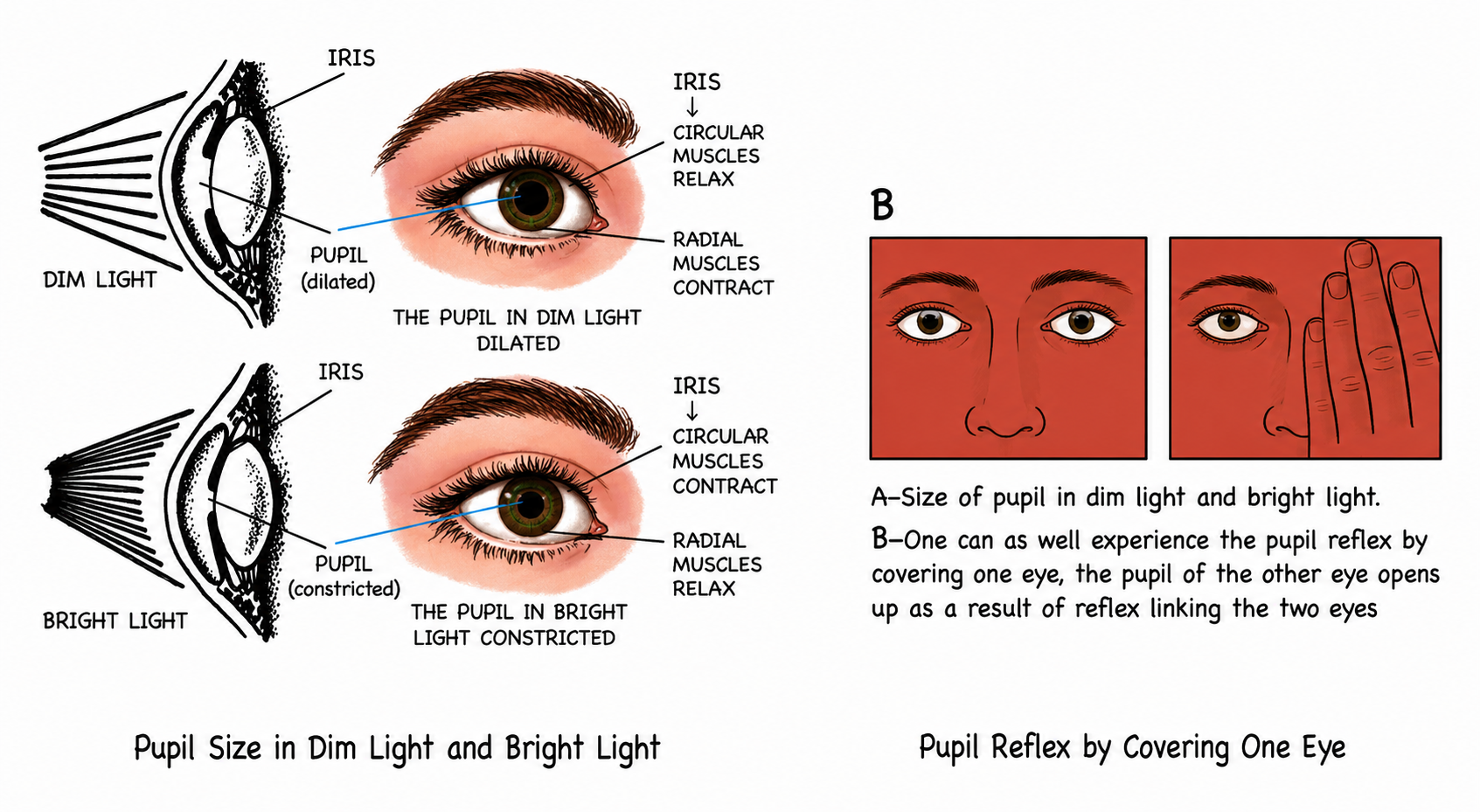
the Pupil. The iris regulates the amount of light entering by dilating or
constricting the pupil.
- Retina: The innermost, light-sensitive layer containing photoreceptor cells.
- Rods: Sensitive to dim light. Contain the pigment
Rhodopsin. Provide black-and-white vision.
- Cones: Sensitive to bright light and colours. Contain the pigment
Iodopsin.
Comparison of rods and cones
| Rods |
Cones |
| 1. More numerous. |
1. Less numerous. |
| 2. Mostly at the periphery of retina. |
2. Mostly located in the centre of retina. |
| 3. Very sensitive to low levels of illumination. |
3. Only stimulated by bright light. |
| 4. One type of rods only, stimulated by most wavelengths of visible light except red. |
4. Three types of cone, each selectively responsive to different wavelengths, therefore, allowing
colour perception. |
| 5. Rapid regeneration of light-sensitive pigment, therefore, can perceive flicker well. |
5. Slower regeneration of light-sensitive pigment, therefore, less responsive to flicker. |
Important Regions on the Retina
- Yellow Spot (Macula Lutea) / Fovea Centralis: Located at the centre of the horizontal
axis of the eye. It contains a very high concentration of cones and NO rods. It is the region of
brightest vision and sharpest image formation.
- Blind Spot: The point where the optic nerve leaves the eye. It contains NO rods and NO
cones, hence no image is formed here.
The Lens and Chambers
- Lens: A transparent, biconvex, flexible crystalline body located just behind the pupil.
It is held in place by Suspensory Ligaments, which are attached to the Ciliary
Body. The ciliary muscles alter the shape of the lens for focusing.
- Aqueous Chamber: The front space between the cornea and lens, filled with watery
Aqueous Humour (keeps the lens moist and protects it from physical shock, refracts
light).
- Vitreous Chamber: The larger space behind the lens, filled with a jelly-like thicker
fluid called
Vitreous Humour (helps in keeping the shape of the eyeball, protects the retina and its
nerve endings).
How do we see?
- Entry of light rays: Light rays from the object enter the eyes through the
transparent structures (conjunctiva, cornea, aqueous humour, lens, vitreous humour).
- Focusing of image: First, the curvature of the cornea converges the light rays to
some extent and the lens converges them further to form an image on the retina. The image on the
retina is inverted and real.
- Nerve impulse produced in retina transmitted to brain: The light energy of the
image produces chemical changes in the sensitive cells (rods and cones). These changes generate
nerve impulses which travel through the optic nerve and reach the visual area of the cerebrum.
- Brain interprets: Our brain interprets the image in many ways, e.g. it "sees" the
objects upright even if the image formed in the eye is inverted.
Accommodation of the Eye
Accommodation: The process of focusing the eye at different distances by changing the
curvature (and thus the focal length) of the lens.
- Viewing Near Objects: Ciliary muscles contract → Suspensory
ligaments relax → Lens becomes thicker and more convex.
- Viewing Distant Objects: Ciliary muscles relax → Suspensory
ligaments become taut/tight → Lens is stretched and becomes thinner/flatter.
Light and Dark Adaptation
When you pass from a brightly lighted area to a dark room (such as a cinema hall), you experience difficulty
in seeing objects for a short while. Slowly, your vision is improved. This improvement is called
Dark Adaptation. This change is due to (a) regeneration of the visual purple (rhodopsin),
the pigment of the rods, which was earlier broken down due to bright light, and (b) dilation of the pupil
permitting more light to enter the eyes.
Conversely, moving from dark to bright light causes a dazzling effect for a short period. After a few
seconds, he comes back to normal viewing through Light Adaptation. The adaptation is due to
reverse of the previous changes, i.e., (a) the visual purple of the rods is bleached, reducing their
sensitivity, and (b) the pupil constricts (gets narrower), to reduce the amount of light entering the eyes.
Colour Vision
Colour vision is possible only through cones of the retina which are stimulated only in bright light. You
cannot make out the red, violet or purple flowers in a garden on a moonlit night, because then only the rods
function and not the cones.
Stereoscopic (binocular) Vision
All monkeys/apes and particularly humans can perceive depth or the relative distance of the objects. This is
due to simultaneous focusing of an object in both eyes, and their images by a kind of "overlapping" in the
brain giving the three dimensional effect.
After-images
If one looks at a bright object for a moment and then closes the eyes, the sensation of light persists
for a short period. In the same way, if one looks at a brightly coloured object and then looks at a dark
surface, an image of the object in the same colour will persist. This is known as persistence
image or the after-image. It lasts for about one-tenth of a second. This
is the principle on which the technique of motion pictures is based.
9.11 Defects of the Eye
- Myopia (Short-sightedness): Can see near objects clearly, but distant objects are
blurred.
- Causes: Eyeball is lengthened from front to back, or the lens is too curved/convex.
- Result: The image is formed in front of the retina.
- Correction: Using a Concave (diverging) lens.
- Hypermetropia (Long-sightedness): Can see distant objects clearly, but near objects
are blurred.
- Causes: Eyeball is shortened from front to back, or the lens is too flat.
- Result: The image is formed behind the retina.
- Correction: Using a Convex (converging) lens.
- Astigmatism: Uneven curvature of the cornea or lens, causing some parts of the
object to be in focus while others are out of focus. Corrected by Cylindrical
lenses.
- Presbyopia: Affects older people. The lens loses its flexibility/elasticity, making
it hard to focus on near objects. Corrected by convex lenses.
- Cataract: The lens turns opaque. Vision is cut off. Corrected by surgical removal
of the lens and replacing it with an artificial lens or using spectacles.
- Night Blindness: Inability to see in dim light due to non-formation of rhodopsin.
Caused by Vitamin A deficiency.
- Colour Blindness: A genetic defect (X-linked) where the person cannot distinguish
between certain colours (mostly red and green).
- Squint: In this defect, the two eyes somewhat converge leading to what is called
"cross eye". An opposite condition appears when they diverge and is called the "wide eye". Corrected
by surgery and suitable exercise.
 and its correction by a CONCAVE LENS..png)
 and its correction by a CONVEX LENS..png)
9.12 Sense Organs: The Ear
The human ear performs two completely different, yet vital functions:
- Hearing
- Body Balance (Equilibrium)
Structure of the Ear
The ear is divided into three main divisions:
1. Outer Ear
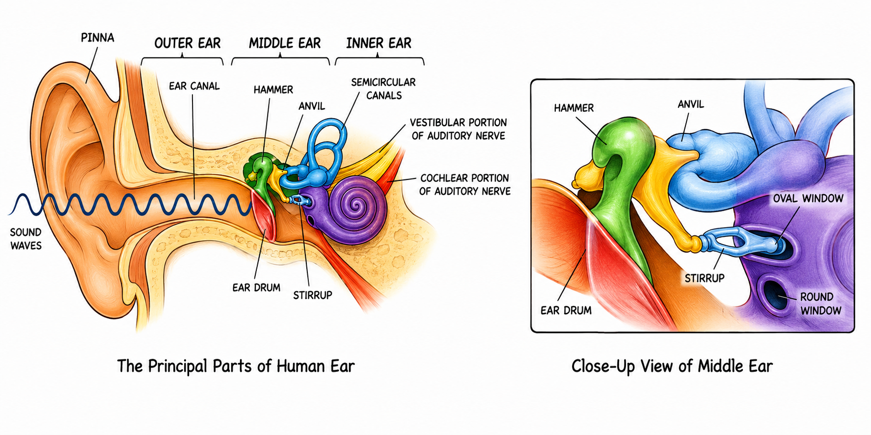
- Pinna (Auricle): The projecting cartilaginous part. It collects and directs sound
waves.
- Auditory Canal: The passage leading inwards. It contains sebaceous glands that
secrete wax (cerumen) to trap dust and insects.
2. Middle Ear
An air-filled cavity containing:
- Tympanum (Eardrum): A tightly stretched membrane that vibrates when struck by sound
waves.
- Ear Ossicles: A chain of three tiny bones: Malleus (Hammer),
Incus (Anvil), and Stapes (Stirrup). They amplify and transmit
vibrations to the inner ear.
- Eustachian Tube: A tube connecting the middle ear to the throat. It equalizes air
pressure on both sides of the eardrum.
- Oval Window (Fenestra ovalis): A membrane-covered opening leading to the inner ear.
The stapes fits into it.
- Round Window (Fenestra rotunda): Another opening covered by a membrane, allowing
for fluid displacement in the cochlea.
3. Inner Ear (Membranous Labyrinth)
The inner ear is a complex system of interconnected tubes and sacs filled with a fluid called
endolymph, surrounded by a bony cavity filled with perilymph. It has
two main parts:
- Cochlea (For Hearing): A snail-shell-like coiled tube. Its central canal contains
the sensory Organ of Corti, which has hair cells that act as hearing receptors. It
connects to the Auditory Nerve.
- Vestibular Apparatus (For Balance): Consists of:
- Three Semicircular Canals: Arranged at right angles to each other. Their
swollen bases (ampullae) contain sensory cells that detect Dynamic Balance
(movement, turning, spinning).
- Utriculus and Sacculus: Sac-like structures that contain sensory cells to
detect Static Balance (gravity, posture when standing still). Connects to
the Vestibular Nerve.

Mechanism of Hearing
Sound waves → Pinna → Auditory Canal → Tympanum (vibrates) → Ear Ossicles (amplify
vibrations) → Oval Window → Cochlear fluid (endolymph/perilymph) → Organ of Corti (hair cells
stimulated) → Auditory Nerve → Brain (Cerebrum) where sound is interpreted.
Mechanism of Balancing
As the head is turned in different directions, the fluid inside the semicircular canals is also shaken. The
moving fluid pushes against sensory hair cells sending the nerve impulse through the auditory nerve to the
brain. The sensory cells in the semicircular canals are concerned with dynamic equilibrium
i.e. while the body is in motion. Similar sensory patches are also located in the utriculus and sacculus
which register the static (positional) balance with respect to gravity.
If you spin round and round, the fluid in the semicircular canals continues to spin for a short time
even after you stop, and you feel dizzy and at the same time, your eyes perform to-and-fro movements
caused due to stimulation of semi-circular canals. Sea-sickness, air-sickness, and
car-sickness are often due to these unusual sensations of equilibrium.
9.13 The Sense of Taste (Gustation)
(Extra - not specified in syllabus)
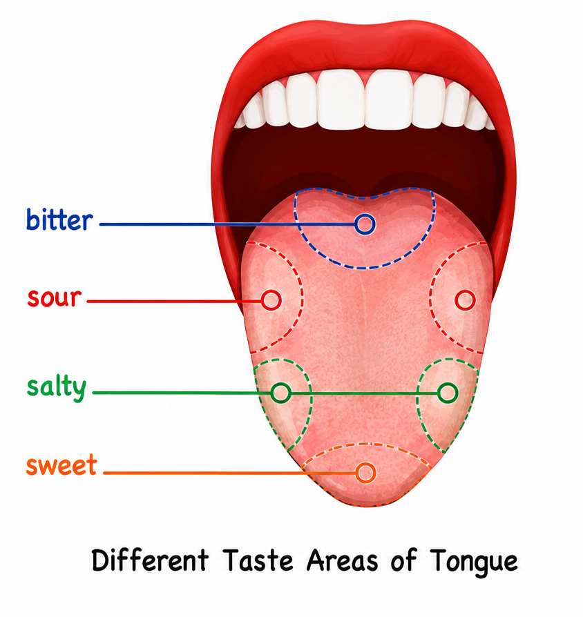
The sense of taste is located in the taste buds of the tongue. The sensory cells end in
hair-like processes and have nerve fibres extending from their bases. Substances in solution enter the taste
pores and stimulate the sensory hairs.
The four fundamental tastes are: Sweet, Salt, Bitter, and Sour.
9.14 The Sense of Smell
(Extra - not specified in syllabus)
The sense of smell is located in the delicate epithelial layers of the nasal chamber. The sensory cells have
hair-like projections which respond to particles dissolved in the mucous secretion of the nose. The impulse
is transmitted to the brain by the olfactory nerve.
Flavour is a combination of taste and smell while eating or sipping. If you hold your
nose, you will find that grated apple and grated onion taste alike - slightly sweet. A cold with a
blocked nose has the same effect and makes food tasteless/flavourless.
Exam Practice Questions (ICSE PYQ Trends)
- NAME THE FOLLOWING The fluid present between the meninges of the brain.
Ans: Cerebrospinal Fluid (CSF).
- NAME THE FOLLOWING The part of the brain that maintains posture and
equilibrium of the body.
Ans: Cerebellum.
- NAME THE FOLLOWING The part of the eye responsible for its shape and
providing a transparent window in the front.
Ans: Sclerotic layer / Cornea.
- NAME THE FOLLOWING The photosensitive pigment present in the rod cells of
the retina.
Ans: Rhodopsin.
- NAME THE FOLLOWING The tube connecting the middle ear to the throat.
Ans: Eustachian Tube.
- NAME THE FOLLOWING The structural and functional unit of the nervous
system.
Ans: Neuron.
REASONING Answer the following:
- Why is the arrangement of white and gray matter different in the brain and spinal
cord?
Ans: In the brain, the cytons (gray matter) are on the outside (cortex) to maximize
surface area via convolutions for higher intelligence. In the spinal cord, axons (white matter) are
on the outside to facilitate rapid conduction of impulses up and down the spinal tracts.
- A person feels dizzy for a moment after spinning around. Why?
Ans: Spinning causes the endolymph fluid inside the semicircular canals to move.
When the spinning stops, the fluid continues to move for a few seconds due to inertia, stimulating
the sensory hair cells and sending false signals of movement to the brain, causing dizziness.
- Why do older people generally require convex lenses for reading?
Ans: Older people suffer from Presbyopia. As age advances, the crystalline lens
loses its elasticity and the ciliary muscles weaken, preventing the lens from becoming convex enough
to focus on near objects. A convex lens corrects this.
APPLICATION Think and answer:
- Two hungry boys (A and B) enter a restaurant and find a table decorated as follows: Boy A
has a raw whole chicken on his plate, Boy B has a roasted chicken. Boy B starts salivating but
not A. Explain the reason for this difference.
Ans: Boy B salivates because of a Conditioned reflex. He has
previous experience and memory of the taste of roasted/cooked food. The sight and smell of the
roasted chicken acts as a stimulus for salivation. Boy A does not salivate because raw chicken is
not associated with a pleasant food memory or experience.
- Identify the part of the autonomic nervous system involved in the following
situations:
| Situation |
Organ |
Change/Action |
Autonomic System |
| You have entered a dark room |
Eye |
Dilation of pupil |
Sympathetic |
| Consuming lot of glucose while running a race |
Liver |
Conversion of glycogen to glucose |
Sympathetic |
| You are chewing a tasty food |
Salivary gland |
Increased secretion of saliva |
Parasympathetic |
| You are running a race |
Adrenal gland |
Secretes adrenaline |
Sympathetic |
| You are retiring to bed for sleep |
Heart |
Heartbeat slows down |
Parasympathetic |
| You are shivering in intense cold |
Body hairs |
Erection of hair |
Sympathetic |
DIFFERENCES Differentiate between the following pairs:
- Myopia and Hypermetropia: In Myopia, the eyeball is too long, the image forms in
front of the retina, and it is corrected by a concave lens. In Hypermetropia, the eyeball is too
short, the image forms behind the retina, and it is corrected by a convex lens.
- Rods and Cones: Rods contain rhodopsin, function in dim light, and provide
black-and-white vision. Cones contain iodopsin, function in bright light, and are responsible for
colour vision.
- Sympathetic and Parasympathetic Nervous System: Sympathetic prepares the body for
stress (fight or flight) by increasing heartbeat and dilating pupils. Parasympathetic restores
normal conditions (rest and digest) by slowing heartbeat and constricting pupils.

, Excited region (Depolarisation), and Recovery region (Repolarise.png)
 apparatus of the human eye showing Lacrimal gland, ducts, sac, and nasolacrimal duct..png)
 of the human eye. (Essential for labelling Cornea, Iris, Lens, Retina, Yellow spot, Blind spot, Optic n.png)