The Circulatory System
ICSE Class 10 Biology • Chapter 07 • Detailed Master Notes
Almost all organisms, including humans, have fluids circulating in their
bodies. Such fluids constitute a distributing system (to supply
substances) as well as a collecting system (to pick up substances) to
and from various parts of the body including the remotest cell.
7.1 Need for Transport
All living cells need continuous supply of nutrients and oxygen, and a
continuous removal of waste products and carbon dioxide. In simple
organisms (like Amoeba), this occurs by simple diffusion. In
multicellular, complex organisms like humans, the cells are located deep
inside the body, hence a specialized
Circulatory System is required.
7.2 Fluids in our Body
There are three principal fluids in our body:
-
Blood: Contained in the heart and in the blood
vessels (arteries, veins, and capillaries) of the circulatory system.
-
Tissue Fluid: Occupies the spaces between cells in
the organs.
-
Lymph: Contained within lymph vessels and lymphatic
organs (like the spleen and tonsils).
Non-circulating fluids: There are also some other
fluids located in particular organs which do not circulate. For
example: synovial fluid filled in the cavities of
skeletal joints, and vitreous humour in the eye.
. Shows relationsh.png)
Fig. 7.1: Diagrammatic representation of blood and lymph circulation
(Red-oxygenated blood, Blue-deoxygenated blood). Shows relationship
between blood, tissue fluid, and lymph.
7.3 Blood
Blood is the most important circulating fluid. It is a slightly thick,
opaque liquid. It is never stationary and is always in
motion from the heart to the arteries and back through the veins.
-
Colour: Bright red (when oxygenated in arteries) and
dark red (when deoxygenated in veins).
- Taste: Slightly salty.
-
pH: Slightly alkaline (pH =
7.3 to 7.4).
-
Volume: An average adult human has about
5 to 6 litres of blood (about 8% of body weight).
7.3.1 Functions of Blood
The functions of blood fall under two main categories:
Transport and Protection.
A. Transport
-
1. Transport of Digested Food: Absorbed in the
alimentary canal (glucose, amino acids) and supplied to all tissues.
-
2. Transport of Oxygen: From the lungs to tissues by
combining with Haemoglobin to form an unstable compound,
Oxyhaemoglobin.
-
3. Transport of Carbon dioxide: From tissues to
lungs, partly dissolved in plasma and partly combined with haemoglobin
as Carbaminohaemoglobin.
-
4. Transport of Excretory Material: Urea and uric
acid are transported from tissues to the liver and kidneys for
elimination.
-
5. Distribution of Hormones: Secreted by endocrine
glands directly into the blood and transported to target organs.
-
6. Distribution of Heat: Helps in regulating body
temperature to a constant $37^\circ C$ by distributing heat uniformly.
B. Protection
-
1. Clotting: Blood forms a clot at the site of injury
to prevent further loss of blood and entry of disease-causing germs.
-
2. Engulfing Bacteria (Phagocytosis): White Blood
Cells (WBCs) protect the body by engulfing and destroying bacteria.
-
3. Antibodies: Blood produces antibodies and
antitoxins which neutralize poisonous substances produced by germs.
7.4 Composition of Blood
Blood consists of two main parts:
- Plasma: The fluid part (about 55%).
-
Cellular Elements: Red Blood Cells, White Blood
Cells, and Platelets (about 45%).
1. Plasma
Plasma is a light yellow, alkaline liquid containing about 90-92% water
and 8% proteins and inorganic salts. It contains
fibrinogen, a protein necessary for blood clotting.
Serum: The plasma from which the protein fibrinogen
has been removed. (Serum = Plasma - Fibrinogen).
2. Red Blood Cells (RBCs or Erythrocytes)
These are the most numerous cells in the blood (about
5 million per cubic mm in adult males and slightly
less, about 4.5 million, in adult females).
-
Shape: Biconcave discs (indented on both sides). They
are very small (about 7 microns in diameter).
-
Site of Production: In adults, produced in the
marrow of long bones (especially ribs, breast bone,
and ilium). In embryos, produced in the liver and spleen. In children,
produced in the bone marrow of all bones until 5 years of age.
-
Small size advantage: Helps them squeeze through the
narrowest blood capillaries in a single file, maximizing the surface
area exposed for oxygen exchange.
-
Haemoglobin: An iron-containing respiratory pigment
present in RBCs that gives blood its red colour and has a high
affinity for oxygen.
Mammalian RBCs lack certain organelles. Why?
ICSE REASONING FAVOURITE Mammalian
RBCs do not have a nucleus, mitochondria, or endoplasmic reticulum
when they mature. This significantly increases their efficiency:
-
Absence of Nucleus: Increases the surface
area/volume ratio for absorbing more oxygen and allows them to carry
more Haemoglobin. It also allows the biconcave shape.
-
Absence of Mitochondria: Mitochondria use up oxygen
for cellular respiration. Without them, RBCs cannot use the oxygen
they are transporting, delivering 100% of it to tissues.
-
Absence of Endoplasmic Reticulum: Increases the
flexibility of RBCs to easily squeeze through extremely narrow
capillaries.
Life Span and Destruction: The average life span of an
RBC is 120 days. Old and weak RBCs are destroyed
primarily in the Spleen, liver, and
bone marrow. The iron part is retained, while the rest is excreted as
bile pigments (bilirubin).
Important facts about RBC count:
-
New born infants have a larger number (6-7 million per cubic mm).
-
RBC count is lower by 5% during sleep, and higher during physical
activity or pregnancy.
-
People living at a height of 4,200m and above have nearly 30% more
RBCs.
-
An abnormally increased number of RBCs is called
Polycythaemia.
-
An abnormally decreased number of RBCs is called
Erythropenia.
Carbon Monoxide Poisoning:
Haemoglobin has a much stronger affinity (about 250 times more) for
Carbon monoxide ($CO$) than for Oxygen. When $CO$ is inhaled, it forms
a highly stable compound called
Carboxyhaemoglobin. This drastically cuts down the
transport of oxygen to tissues, often leading to death by
asphyxiation.
3. White Blood Cells (WBCs or Leukocytes)
WBCs are far fewer in number (about 4000 to 8000 per cubic mm) and lack
haemoglobin.
-
Shape: They are mostly amoeboid (can change shape)
and possess a distinct nucleus.
Diapedesis: The process by which White Blood Cells
(especially neutrophils) squeeze out through the walls of the blood
capillaries to the site of infection.
Phagocytosis: The process by which WBCs engulf and
destroy disease-causing pathogens or foreign particles.
 and Phagocytosis (engulfing bacteria)..png)
Fig. 7.13: Diagrammatic representation of Diapedesis (WBCs squeezing
through capillary walls) and Phagocytosis (engulfing bacteria).
Categories of WBCs
| Type |
Sub-types |
Key Characteristics & Functions |
|
Granular WBCs (Have granular cytoplasm and lobed
nucleus)
|
1. Neutrophils
2.
Eosinophils
3. Basophils
|
1. Phagocytosis (main cellular defence).
2. Associated with
allergy and parasitic infections.
3. Secrete histamine for
inflammation.
|
|
Agranular WBCs (Have clear cytoplasm and single
large nucleus)
|
1. Lymphocytes
2. Monocytes
|
1. Produce Antibodies.
2. Actively
phagocytic (can transform into macrophages).
|
4. Blood Platelets (Thrombocytes)
These are very minute, oval, non-nucleated fragments of giant cells
(megakaryocytes) formed in the red bone marrow. They number about
200,000 to 400,000 per cubic mm. They are essential for
blood clotting.
Note: If the number of platelets falls to an abnormally low
count, coagulation occurs very slowly and often leads to haemorrhage.
This occurs in viral dengue fever.
7.5 Coagulation of Blood (Blood Clotting)
When a blood vessel is cut, blood escapes, but soon a clot is formed
preventing further loss. This cascade mechanism involves several steps:
-
Injured tissue cells and the platelets disintegrate and release an
enzyme called Thrombokinase (also called
Thromboplastin, or Factor X / Stuart factor).
-
Thrombokinase acts as an enzyme and, with the help of
Calcium ions ($Ca^{++}$) present
in the plasma, converts the inactive protein
Prothrombin into the active enzyme
Thrombin.
-
Thrombin reacts with the soluble plasma protein
Fibrinogen, converting it into insoluble
Fibrin.
-
Fibrin forms a sticky mesh or network of threads at the wound site.
RBCs get trapped in this mesh to form the solid
Blood Clot (Thrombus).
Haemophilia: A genetic disorder where the blood does not clot
normally due to a lack of certain clotting factors.
7.6 Blood Groups and Transfusion
Blood is not entirely identical in all humans. It is classified under
the ABO system based on the presence or absence of
specific proteins on the RBC surface called Antigens (A
and B), and complementary proteins in the plasma called
Antibodies (anti-A and anti-B).
| Blood Group |
Antigen on RBC |
Antibody in Plasma |
Can Donate To |
Can Receive From |
| A |
A |
Anti-B |
A, AB |
A, O |
| B |
B |
Anti-A |
B, AB |
B, O |
| AB (Universal Recipient) |
A and B |
None |
AB only |
All (A, B, AB, O) |
| O (Universal Donor) |
None |
Anti-A and Anti-B |
All (A, B, AB, O) |
O only |
The Rh Factor
Besides ABO antigens, another antigen called the Rh factor (Rhesus
factor) may be present. People with this antigen are
Rh positive (Rh+), and those without it are
Rh negative (Rh-).
Pregnancy Complication: If an Rh- mother carries an Rh+ fetus,
the mother's blood may develop antibodies against the Rh+ cells. While
the first child is usually safe, subsequent Rh+ pregnancies can result
in the destruction of fetal RBCs, a severe condition called
erythroblastosis fetalis.
7.7 The Blood Vessels
Blood flows through three main types of vessels in a closed circuit.
-
Arteries: Vessels that carry blood
away from the heart to any organ.
-
They have thick, highly muscular, and elastic walls to withstand
the high pressure of blood pumped by the heart.
- They have a narrow lumen and no valves.
-
They carry oxygenated blood (except the Pulmonary Artery).
-
Veins: Vessels that carry blood
towards the heart from the organs.
-
They have thin walls and a wider lumen as blood flows under low
pressure.
-
They possess pocket-shaped valves to prevent
the backflow of blood.
-
They carry deoxygenated blood (except the Pulmonary Vein).
-
Capillaries: The ultimate extremely fine branches
of arterioles.
-
Their walls consist of a
single layer of squamous epithelial cells
(endothelium).
-
They allow the exchange of oxygen, carbon dioxide, dissolved
nutrients, and excretory products between the blood and tissues.
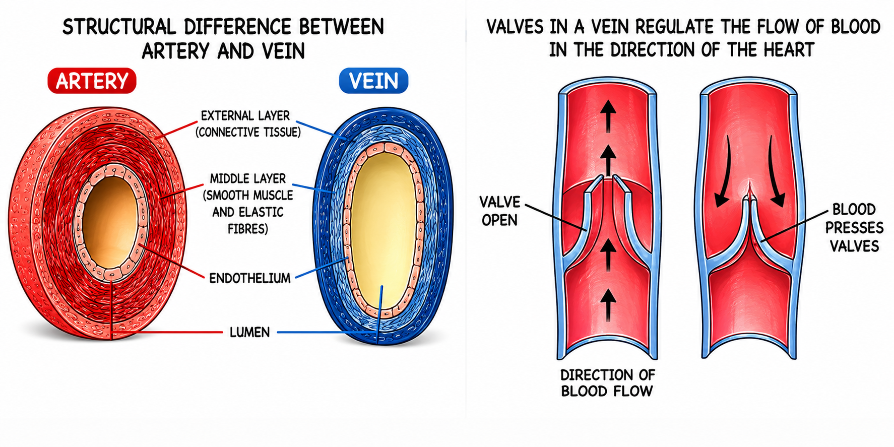
Fig. 7.10 & 7.11: Structural difference between artery and vein, and
valves in a vein regulating the flow of blood in the direction of the
heart.
7.8 The Heart
The heart is a muscular pumping organ located in the centre of the chest
cavity, slightly tilted to the left. It is about the size of one's
closed fist.
-
Protective Covering: The heart is enclosed by a
double-walled membranous sac called the
Pericardium. The space between the
two membranes is filled with pericardial fluid, which reduces friction
and protects the heart from mechanical shocks.
Chambers of the Heart
The mammalian heart has four chambers:
-
Two Auricles (Atria): The upper chambers. They have
thin walls because their major function is only to receive blood and
pump it into the ventricles directly below them.
-
Two Ventricles: The lower chambers. They have thick,
muscular walls because they have to pump blood over long distances.
Note: The wall of the
Left Ventricle is much thicker than the right
ventricle because it has to pump blood to the farthest extremities of
the entire body, whereas the right ventricle only pumps blood a short
distance to the lungs.
Valves of the Heart
Valves are flap-like structures that allow blood to flow in only one
direction.
-
Right Atrioventricular Valve (Tricuspid Valve):
Located between the right auricle and right ventricle. It has three
flaps (cusps).
-
Left Atrioventricular Valve (Bicuspid or Mitral Valve):
Located between the left auricle and left ventricle. It has two flaps.
-
Semilunar Valves: Pocket-shaped valves located at the
origin of the Pulmonary Artery and the Aorta to prevent backflow into
the ventricles.
Chordae Tendineae: Tough, inelastic tendinous cords
that attach the flaps of the tricuspid and bicuspid valves to the
papillary muscles of the ventricle walls. They prevent the valves from
turning inside out like an umbrella during strong ventricular
contractions.
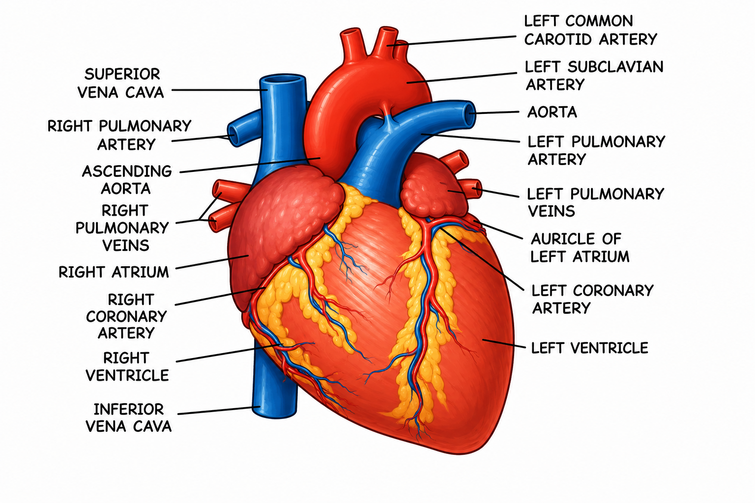
Fig. 7.8: Internal Structure of the Human Heart.
(Master this diagram for labeling: Vena Cava, Aorta, Pulmonary
Artery/Vein, Auricles, Ventricles, Valves, and Chordae
tendineae).
7.9 Circulation of Blood in the Heart (Double Circulation)
Blood flows twice through the heart to complete one full cycle. This is
called Double Circulation.
-
Pulmonary Circulation: The flow of deoxygenated blood
from the right ventricle to the lungs (via pulmonary artery) for
oxygenation, and the return of oxygenated blood to the left auricle
(via pulmonary veins).
-
Systemic Circulation: The flow of oxygenated blood
from the left ventricle to all body parts (via aorta), and the return
of deoxygenated blood to the right auricle (via vena cavae).
 and systemic circuit (bottom)..png)
Fig. 7.10: Diagrammatic representation of Double Circulation showing
the pulmonary circuit (top) and systemic circuit (bottom).
7.10 Hepatic Portal System
Portal System: A system where a vein starts with
capillaries in one organ and ends in capillaries in another organ,
instead of going directly to the heart.
The Hepatic Portal Vein collects nutrient-rich blood
from the stomach and intestines and enters the Liver,
where it breaks up into capillaries.
Importance: The liver acts as a checkpoint. It extracts excess
glucose and stores it as glycogen, detoxifies harmful substances, and
processes amino acids before the blood enters the general circulation.
7.11 Heart Beat and Cardiac Cycle
A complete heartbeat consists of the contraction (Systole) and
relaxation (Diastole) of the heart chambers.
-
Sino-atrial Node (SAN) or Pacemaker: A specialized
patch of tissue in the wall of the right auricle that originates the
electrical impulse for the heartbeat.
-
The impulse spreads to the
Atrio-ventricular Node (AVN), then travels down the
Bundle of His, and finally into the ventricle walls
via the Purkinje fibres, causing the ventricles to
contract.
Heart Sounds
You can hear two distinct sounds through a stethoscope during a
heartbeat:
-
LUBB (First sound): Produced during ventricular
systole when the tricuspid and bicuspid (AV) valves close sharply to
prevent backflow.
-
DUP (Second sound): Produced at the beginning of
ventricular diastole when the semilunar valves at the roots of the
aorta and pulmonary artery close.
7.12 Blood Pressure and Pulse
-
Pulse: The alternate expansion and elastic recoil of
the wall of the artery during ventricular systole. The arteries
distend due to their elastic muscular walls. This distension can be
easily felt by pressing gently over a superficial artery, such as the
radial artery of the wrist. Counting the pulse is
indirectly counting the heart beat. The pulse rate increases due to
physical exercise or certain emotions.
-
Blood Pressure: The pressure which the blood flowing
through the arteries exerts on their walls. It has two limits:
-
Systolic Pressure (Upper limit): Recorded when
fresh blood is pushing through the artery as a result of
ventricular contraction. Normal range:
100 - 140 mm Hg.
-
Diastolic Pressure (Lower limit): Recorded when
the wave has passed over (ventricular relaxation). Normal range:
60 - 80 mm Hg.
Blood pressure is measured using a Sphygmomanometer.
A rise in blood pressure above
140/90 is known as
Hypertension (high blood pressure).
7.13 Tissue Fluid and Lymphatic System
As blood flows under high pressure in the capillaries, the liquid part
(plasma minus large proteins) and some WBCs (by diapedesis) leak out
through the walls and bathe the cells. This fluid is called
Tissue Fluid. It helps in the actual exchange of
materials directly with the cells.
Most of this fluid re-enters the blood capillaries, but some enters
another set of minute channels named lymph vessels, where it is called
Lymph. The lymph flows in these vessels due to the
contraction of surrounding muscles (a beneficial effect of physical
exercise) and ultimately pours back into major anterior veins close to
the right auricle, returning to circulation.
Composition of Lymph
-
Cellular part: Only leukocytes (mostly lymphocytes).
No blood platelets.
-
Non-cellular part: Water (94%) and Solids (6% -
proteins, fats, carbohydrates, enzymes, antibodies, etc.).
Functions of Lymph
-
Nutritive: Supplies nutrition and oxygen to those
parts where blood cannot reach.
-
Drainage: Drains away excess tissue fluid and
metabolites, returning proteins to the blood from tissue spaces.
-
Absorption: Fats from the intestine are absorbed
through lymphatics called lacteals located in the
intestinal villi.
-
Defence: Lymphocytes and monocytes of the lymph
function to defend the body. Lymph nodes filter out and destroy
bacteria.
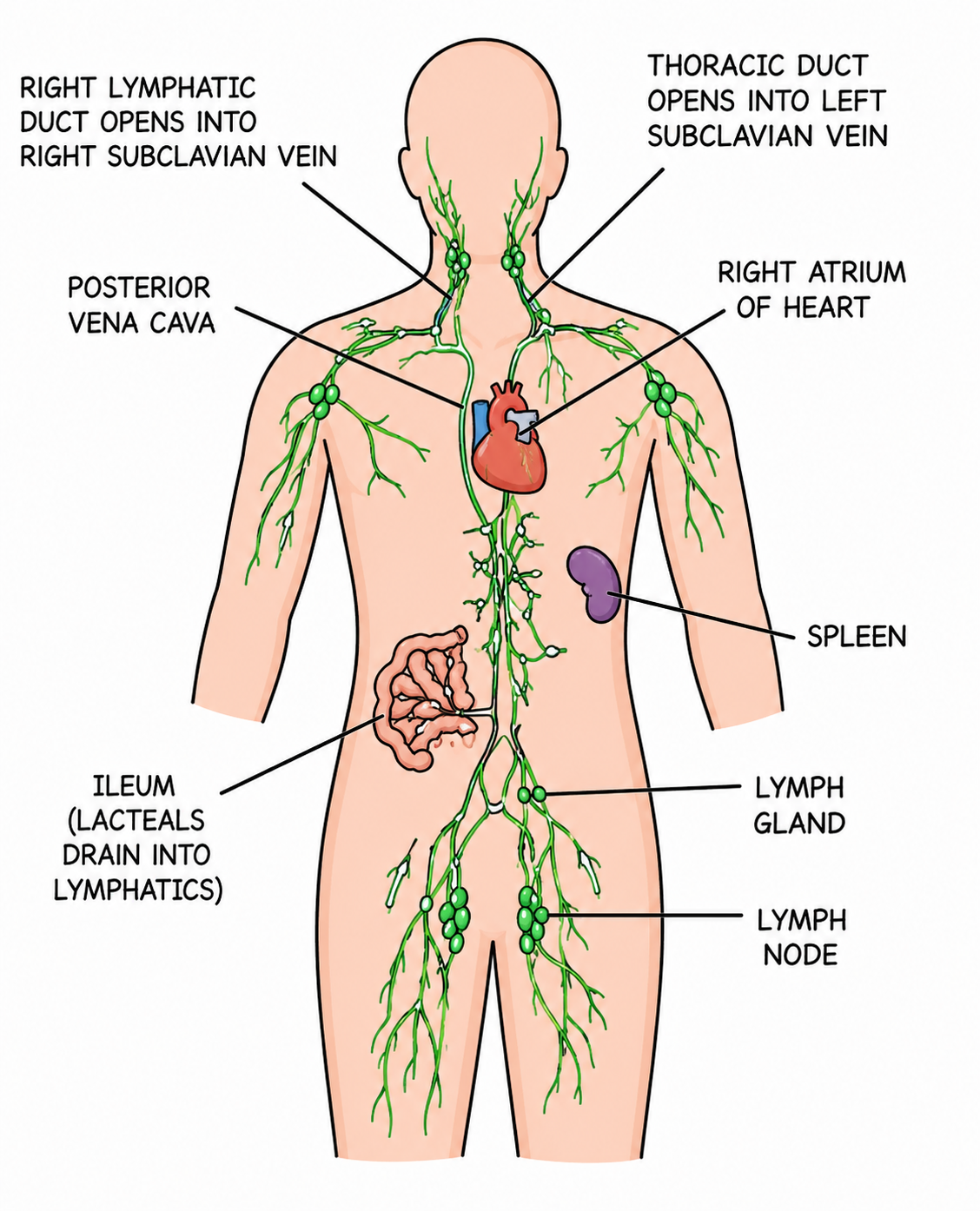
Fig. 7.16: Distribution of main lymph vessels in the human body.
(Shows Right Lymphatic Duct, Thoracic Duct, Spleen, Lymph Nodes, and
Lymph Gland).
The Spleen
A large lymphatic organ situated in the abdomen behind the stomach.
Functions:
- Acts as a blood reservoir.
- Produces lymphocytes.
-
Destroys old and worn-out RBCs (known as the "Graveyard of RBCs").
- In embryos, it produces RBCs.
Exam Practice Questions (ICSE PYQ Trends)
-
NAME THE FOLLOWING The blood vessel that
supplies oxygenated blood to the heart muscles.
Ans:
Coronary Artery
-
NAME THE FOLLOWING The protective double
membranous covering of the heart.
Ans:
Pericardium
-
NAME THE FOLLOWING The process of WBCs
squeezing out through the walls of blood capillaries.
Ans:
Diapedesis
-
NAME THE FOLLOWING The mineral element
essential for the clotting of blood.
Ans:
Calcium ($Ca^{++}$)
REASONING Answer the following:
-
Why do mature mammalian RBCs lack a nucleus and
mitochondria?
Ans: The absence of a nucleus provides more surface
area to pack haemoglobin and gives the biconcave shape. The absence
of mitochondria ensures that the RBC does not use the oxygen it
transports for its own respiration, delivering it fully to the
tissues.
-
Why does the left ventricle have thicker muscular walls than the
right ventricle?
Ans: The left ventricle has to pump blood under
extremely high pressure to the farthest extremities of the entire
body, whereas the right ventricle only has to pump blood a short
distance to the nearby lungs.
-
Why do veins have valves while arteries do not?
Ans: Veins carry blood at low pressure against
gravity, so pocket valves are required to prevent the backflow of
blood. Arteries carry blood at high pressure direct from the heart,
making backflow impossible.
DIFFERENCES Differentiate between the
following pairs based on criteria given in brackets:
-
Artery and Vein (Direction of blood flow): Artery
carries blood away from the heart. Vein carries blood towards the
heart.
-
LUBB and DUP (Valves involved): LUBB is produced by
the closure of the Atrioventricular (Bicuspid and Tricuspid) valves.
DUP is produced by the closure of the Semilunar valves at the roots
of the aorta and pulmonary artery.
-
Blood and Lymph (Composition): Blood contains RBCs,
WBCs, Platelets, and plasma. Lymph contains only WBCs (mainly
lymphocytes) and plasma without large proteins (no RBCs).
PRACTICE Try these textbook review
questions:
-
Which valve is present between the right atrium and the right
ventricle?
Ans: Tricuspid valve
-
The blood vessel supplying blood to the kidney is the:
Ans: Renal artery
-
Heart sounds are produced due to:
Ans: Closure of atrioventricular and semilunar
valves.
-
Name the kind of blood cells which can squeeze out through the
walls of one category of blood vessels:
Ans: White Blood Cells (Leukocytes) via diapedesis.
-
Name an artery which carries impure (deoxygenated) blood:
Ans: Pulmonary artery