Structure of Chromosomes, Cell Cycle and Cell Division
ICSE Class 10 Biology Detailed Master Notes (Chapter 2)
Cell division is one of the most fundamental characteristics of life.
This is the method which enables life to perpetuate generation after
generation. It is equally true for simple organisms like Amoeba as it is
for complex ones like humans or giant elephants.
1. Structure of Chromosomes
The most conspicuous events occurring during cell division are all
related to chromosomes. The duplicated chromosomes get evenly
distributed into daughter cells during mitosis, ensuring normal
functioning of the cells.
1.1 What are Chromosomes?
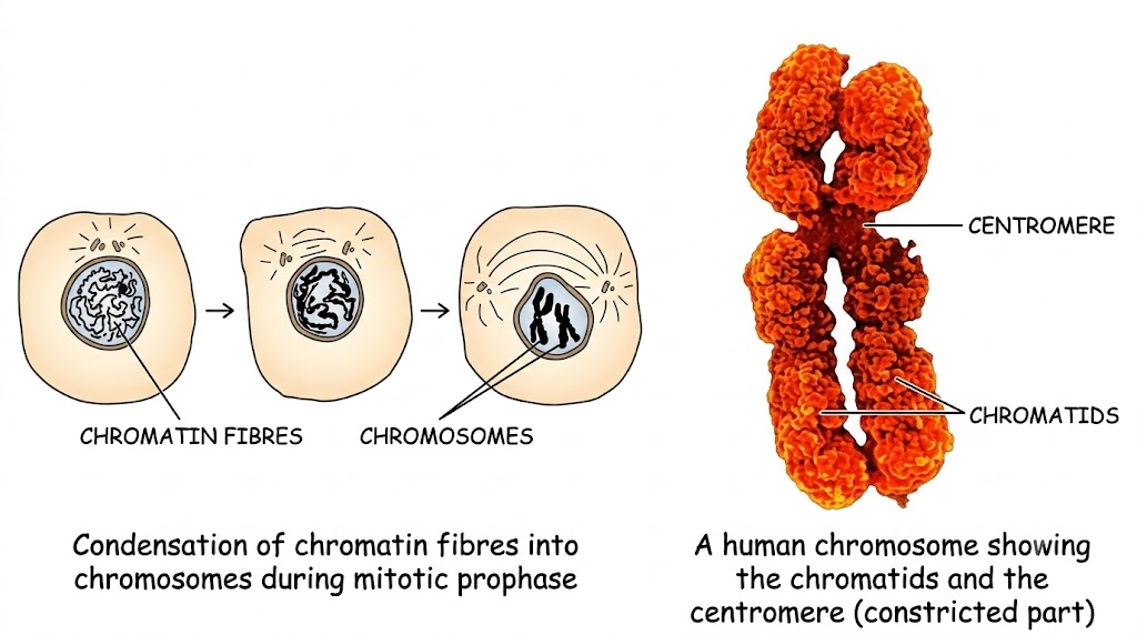
Chromosomes are highly condensed, coiled
chromatin fibres.
-
When a normal, unstained living cell is observed under a light
microscope, its nucleus may appear empty.
-
When stained with suitable dyes, structures become noticeable. In the
interphase (non-dividing stage), the nucleus shows a
network of very long, extremely thin dark-staining fibres called
chromatin fibres.
-
As the cell enters the first stage of division (prophase), the
chromatin fibres condense to form chromosomes.
-
The name comes from their ability to pick up dyes easily:
chroma = coloured, soma = body.
1.2 Discovery of Chromosomes
-
Discovered in 1882 by German Scientist
Walther Fleming.
-
He noticed them in the rapidly dividing cells of salamander larvae (an
amphibian).
-
Using an old-type microscope, he saw minute threads appearing to
divide lengthwise. He coined the term
mitosis (literally meaning
"thread").
1.3 Gross Structure of a Chromosome
Each chromosome, in its condensed form at the start of cell division,
consists of:
-
Two Chromatids: The two identical halves of a
duplicated chromosome.
-
Centromere: The point of attachment where the two
sister chromatids are joined. It appears as a small constricted
region. The centromere is also the exact site that attaches to the
spindle fibre during cell division.
As the spindle fibre contracts, the sister chromatids are separated at
the centromere and pulled towards opposite poles. After division,
chromatids (now individual chromosomes) decondense back into chromatin
fibres.
Chemical Composition of Chromatin
The chromatin material is composed of two primary substances:
-
DNA (Deoxyribonucleic acid): Constitutes about
40%.
-
Histones: A particular type of basic protein
constituting about 60%.
1.4 Molecular Structure (The Nucleosome & DNA)
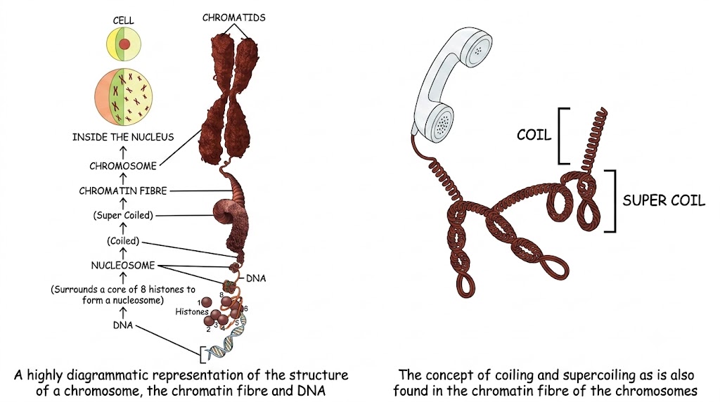
The Nucleosome: The DNA strand winds around a core of
exactly eight histone molecules. This core can be
imagined like a football around which a long rope is wound with one or
two loops. Each such complex is called a
nucleosome.
-
A single human chromosome may contain about a million nucleosomes.
-
The entire chromatin fibre undergoes extensive
coiling and supercoiling (similar to a typical
telephone cord).
Structure of DNA
(Note: Shape studied by Rosalind Franklin in 1953; structure worked
out by Watson and Crick in 1953.)
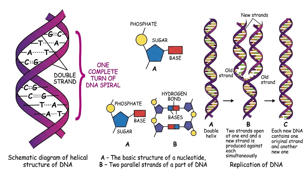
DNA is a very large single molecule, described as a
macromolecule. It is composed of two complementary
strands wound around each other in a
double helix.
Each single DNA strand is composed of repeating structural units called
Nucleotides. A nucleotide consists of three components:
- Phosphate
- Sugar (pentose)
-
Nitrogenous Base (attached to the sugar inwards)
The two strands make a ladder-like arrangement. The alternating sugar
and phosphate form the uprights, and the nitrogenous bases form the
"rungs" by joining via Hydrogen Bonds.
Base Pairing Rules in DNA:
-
Adenine (A) always pairs with
Thymine (T) with
two hydrogen bonds.
-
Guanine (G) always pairs with
Cytosine (C) with
three hydrogen bonds.
Replication of DNA
During the interphase of the cell cycle, DNA duplicates (replicates) to
prepare for equitable distribution during mitosis. The DNA double helix
unzips (opens) at one end, exposing free strands. New strands are built
against each original strand simultaneously. The result is two identical
DNA molecules, each containing one "old" strand and one "new" strand.
1.5 What are Genes?
Genes are specific sequences of nucleotides on a
chromosome that encode particular proteins which express in the form
of some particular feature of the body.
Extra Knowledge (Not in Syllabus but helpful):
DNA Profiling (Fingerprinting): About 99% of DNA is
non-functional and shows tremendous variations from person to person.
This helps in identifying individuals (e.g., establishing paternity or
solving crimes).
Stem Cells: Undifferentiated cells that can undergo
unlimited divisions and give rise to various specialized cells (like
nerve or blood cells).
2. Cell Division - New Cells from Existing Ones
New cells are required in the body for four primary reasons:
-
For Growth: Every organism begins life as a single
cell (the fertilized egg). This cell divides repeatedly to form a
cluster, differentiating into tissues and organs.
-
For Replacement: Wear and tear of cells occurs daily.
Example:
20 million red blood cells are
destroyed every minute in our body and are constantly replaced by new
cells from the bone marrow.
-
For Repair: Cells divide to cover up gaps from
accidental injuries, cuts in the skin, or fractures in bones.
-
For Reproduction: Unicellular organisms (Amoeba,
yeast) divide to form independent cells. In higher forms, special
cells undergo meiosis to produce gametes (sperms and eggs).
Fascinating Cell Longevity Facts:
-
Cells lasting a lifetime (not replaced if dead):
Eye lens cells, nerve cells of the cerebral cortex, and most muscle
cells.
- Skin cells: Replaced every two weeks.
- Red blood cells: Last for about 120 days.
-
Bone cells: Replaced every 10 years in adults.
- Liver cells: Replaced every 300-500 days.
-
Epithelial cells lining the gut: Last only 5 days.
3. Types of Cell Division
There are two primary types of cell division:
-
Mitosis: Cell division leading to growth, repair, and
replacement. Occurs in somatic (body) cells.
-
Meiosis: Cell division leading to the production of
gametes (sex cells). Occurs in reproductive organs.
3.1 Mitosis (mitos: thread)
Mitosis is the cell division in which one parent cell
divides into two identical daughter cells, maintaining the exact same
normal chromosome number (diploid, 2n) at each division.
Before division, the cell prepares by doubling the quantity of DNA
during the Interphase (the apparently resting phase,
though internally very active).
Phases of Mitosis
Mitosis is broadly divided into Karyokinesis (division
of the nucleus) and Cytokinesis (division of the
cytoplasm). Karyokinesis occurs in four continuous phases:
(i) Prophase (pro: first)
- Chromosomes condense, becoming short, thick, and distinct.
-
Each chromosome is already duplicated as paired sister
chromatids attached at the
centromere.
-
In animal cells, the centrosome splits, and
duplicate centrioles move apart to opposite poles.
-
Each centriole is surrounded by radiating microtubules termed an
aster.
-
Spindle fibres appear between the daughter
centrioles.
- The nuclear membrane and nucleolus disappear.
- Chromosomes begin moving toward the cell equator.
(ii) Metaphase (meta: after)
-
The duplicated chromosomes line up along the
equatorial plane of the cell.
-
Each chromosome gets attached to a spindle fibre precisely by its
centromere.
(iii) Anaphase (ana: up, back)
- The centromere dividing the two chromatids finally splits.
-
The two sister chromatids separate and are drawn apart toward
opposite poles.
-
The pulling action is due to the contraction (shortening) of spindle
fibres.
-
In animal cells, a cleavage furrow begins to form in the cell
membrane at the middle.
(iv) Telophase (telo: end)
-
Daughter chromosomes reach opposite poles and thin out, reverting to
a network of chromatin threads.
- Spindle fibres disappear.
- Nuclear membrane and nucleoli reappear.
- In animal cells, the cleavage furrow deepens.
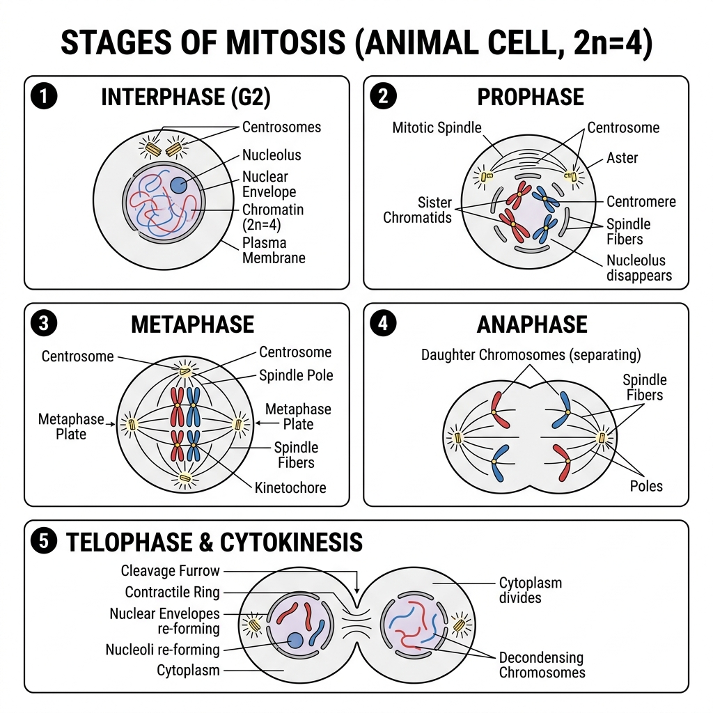
Cytokinesis (Division of Cytoplasm)
At the end of telophase, cytokinesis completes the process. In
animal cells, the cleavage furrow deepens totally and
splits the cell into two. In plant cells, a
cell plate is laid down in the
cytoplasm at the equatorial plane, growing from the center to the
periphery.
Differences between Mitosis in Animal and Plant Cells
| Feature |
Animal Cell Mitosis |
Plant Cell Mitosis |
| Asters |
Asters are formed around centrioles. |
Asters are not formed (no centrosome). |
| Cytokinesis |
By furrowing of the cytoplasm. |
By cell plate formation. |
| Location of occurrence |
Occurs in most tissues throughout the body (for growth and
replacement).
|
Occurs mainly at growing tips (meristems) for lengthening and sides
for increasing girth.
|
Significance of Mitosis
-
Growth: Increase in body size due to new cell
formation.
-
Repair: Healing of damaged and wounded tissues.
-
Replacement: Replacing old/dead cells (e.g., blood
cells, epidermal cells).
-
Asexual Reproduction: Used by unicellular organisms
like Amoeba to divide into two.
-
Genetic Stability: Maintains the exact same
chromosome number in daughter cells.
Mitochondria and Chloroplasts in Cell Division
-
Both possess their own DNA and Ribosomes (which produce specific
proteins).
-
They divide on their own by
simple fission (splitting into two) and are
partitioned between the two daughter cells during mitosis.
4. The Cell Cycle - "Divide, grow and redivide"
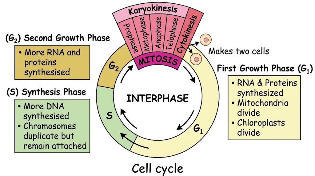
Cell Cycle: The entire sequence of events that a cell
passes through from its formation to its next division.
The interphase prepares the cell for division and consists of three
distinct phases:
-
First Growth Phase ($G_1$): RNA and proteins are
synthesised, the volume of cytoplasm increases. Mitochondria and
chloroplasts divide.
-
Synthesis Phase ($S$): More DNA is synthesised. The
chromosomes strictly duplicate but remain attached at the centromere.
-
Second Growth Phase ($G_2$): A shorter growth phase
where RNA and proteins necessary for cell division continue to be
synthesised.
In late $G_1$ phase, cells may withdraw from the cycle and enter a
Resting phase (R), or proceed to the S phase.
Can the cell cycle go on endlessly?
No. It stops permanently in some cells (like nerve cells), temporarily
in others (like liver cells dividing once every 1-2 years), and runs
continuously in others (skin surface cells, plant meristems).
Uncontrolled, non-stop cell cycles lead to tumours/cancer.
5. Meiosis (Reduction Division)
Meiosis occurs in the reproductive organs (testis and ovary in humans,
anthers and ovary in plants) to produce gametes.
The most significant aspect of meiosis is that the chromosome number is
halved. From 23 pairs (diploid, 2n) in human body
cells, only single chromosomes (haploid, n) are passed to sperms/eggs.
Upon fertilization, the diploid number ($n + n = 2n$) is restored.
Significance of Meiosis
-
Halves Chromosome Number: Essential so fertilization
restores the normal double (diploid) number.
-
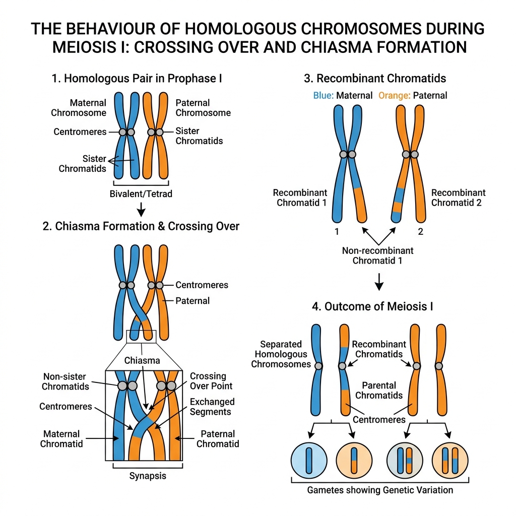
Mixing of Genes (Variations): Occurs in two ways:
-
Maternal and paternal chromosomes mix up randomly as they separate
from homologous pairs.
-
Crossing Over: While separating, chromatid
material often gets exchanged between members of a homologous pair
at a point called the chiasma.
This results in genetic recombination and variation in progeny.
Difference Between Mitosis and Meiosis
| Feature |
Mitosis |
Meiosis |
| 1. Where it occurs |
In the somatic (body) cells. |
In reproductive cells. |
| 2. What for |
Growth, repair, and replacement. |
Only for gamete formation. |
| 3. When it occurs |
Continuously throughout life. |
Only in reproductively active age. |
| 4. Number of daughter cells |
Two daughter cells. |
Four daughter cells. |
| 5. Chromosome number |
Full set (Diploid, 2n). |
Half the number (Haploid, n). |
| 6. Number of nuclear divisions |
A single nuclear division. |
Two nuclear divisions. |
| 7. Identity of genes |
Identical to the parent cell. |
Randomly assorted (results in variations). |
6. ICSE Board Exam Practice Questions
Mastering these questions directly from the syllabus exercises
guarantees you are fully prepared!
-
The chromatin material is formed of:
Ans: (b) DNA and Histones
-
The term "chromosomes" literally means:
Ans: (c) Coloured bodies
-
The number of chromosomes in a certain type of cell division is
halved. This kind of division occurs in:
Ans: (c) both ovary and testis
-
In which option are the two stages of mitosis in correct
sequence?
Ans: (a) Prophase, anaphase
(Since prophase comes before anaphase)
-
Synthesis phase in the cell cycle is called so for the synthesis
of more of:
Ans: (c) DNA
1. Name the following:
-
The repeating components of each DNA strand lengthwise:
Nucleotides
-
The complex structure consisting of DNA strand and a core of
histones: Nucleosome
-
The type of bond which joins the complementary nitrogenous bases:
Hydrogen bond
-
The three components of a nucleotide:
Phosphate, Sugar (pentose), and Nitrogenous base
2. True or False with Reason:
-
As you grow from childhood to adulthood, your skin cells divide
only to replace cells lost from the surface.
False. Skin cells divide for both overall body
growth (increasing surface area) AND replacing lost cells.
-
The unfertilised human egg has half the number of chromosomes of
the body cells.
True. Gametes are produced by meiosis, resulting in
a haploid (n) number of chromosomes.
-
Nuclear membrane in a mitotically dividing cell remains intact up
to the metaphase and disappears only in the telophase.
False. The nuclear membrane disappears entirely by
the end of late prophase.
-
Mitotic cell division can be a mode of reproduction.
True. In unicellular organisms like Amoeba, mitosis
serves as a method of asexual reproduction.
-
Crossing-over between chromatids can occur only between
homologous chromosomes.
True. Crossing over strictly occurs between the
non-sister chromatids of a homologous maternal and paternal pair.
3. Fill in the blanks:
-
(a) Mitosis occurs in our somatic (body) cells.
-
(b) Mitosis produces two daughter cells whereas meiosis produces
four daughter cells.
-
(c) Meiosis occurs only in reproductive cells.
-
(d) Modern humans have 46 chromosomes. Their sperms and eggs will
have 23 chromosomes each.
-
(e) During the pairing of chromosomes in meiosis, the
homologous chromosomes come to lie side by side.
-
(f) The centriole(s) are surrounded by radiating
rays and termed as aster.
1. State the difference between:
-
(a) Chromosome and chromatid: A chromosome is a
highly condensed chromatin fibre. A chromatid is one of the two
identical sister halves of a duplicated chromosome joined at the
centromere.
-
(b) Centrosome and centromere: A centrosome is an
organelle located near the nucleus in animal cells containing
centrioles that initiate cell division. A centromere is the
constricted point on a chromosome where two sister chromatids
attach.
-
(c) Aster and spindle fibres: An aster is a
star-like structure formed of radiating microtubules surrounding a
centriole at the pole. Spindle fibres are the fine threads that run
across the cell between the two poles/asters to which chromosomes
attach.
-
(d) Haploid and diploid: Haploid (n) refers to a
single set of chromosomes (found in gametes). Diploid (2n) refers to
paired sets of chromosomes (found in somatic cells).
2. "First meiotic division is the reduction division". What does
'reduction' refer to?
It refers to the reduction of the chromosome number from diploid (2n)
to haploid (n) as homologous pairs separate.
3. "Gametes must be produced by meiosis for sexual reproduction".
Why?
If gametes were produced by mitosis, they would have a diploid (2n)
number. Fertilization ($2n + 2n$) would result in a $4n$ zygote,
doubling the chromosomes every generation. Meiosis ensures gametes are
haploid ($n$), so fertilization restores the constant $2n$ chromosome
number for the species.
ESSENTIAL FOR SECTION B Carefully analyze
all visual diagrams as they are guaranteed to appear in ICSE.
Scenario 1: Cell A undergoes one mitotic division.
Cell B undergoes one complete meiotic division. How many cells are
produced?
Ans: Cell A produces 2 cells. Cell B produces 4
cells.
Scenario 2: Identify the Stages
-
Chromosomes arranged in a horizontal plane at the equator
$\rightarrow$ Metaphase
-
Daughter chromosomes move to opposite poles $\rightarrow$
Anaphase
-
Chromosomes become visible as fine long threads $\rightarrow$
Prophase
-
Chromosomes lose distinctiveness and turn to chromatin network
$\rightarrow$ Telophase