Vardaan Learning Institute
Life Processes
Definition: The basic maintenance functions performed by living organisms to sustain life are called Life Processes: Nutrition, Respiration, Transportation, Excretion.
Why do we need Life Processes even when at rest or sleeping?
Living organisms are made of carbon-based molecules which are complex and highly organized. These structures tend to break down over time. Even during sleep, the body constantly repairs and maintains its cells using molecular movements — which requires a continuous supply of energy. Without these processes, the organized structure of life would fall apart.
Why is diffusion insufficient for multicellular organisms?
In single-celled organisms (like Amoeba), the entire body surface is in contact with the environment — diffusion is enough for exchange of gases and nutrients.
In multicellular organisms (like humans), cells deep inside the body are NOT in direct contact with the environment. Simple diffusion cannot reach all cells. Hence, specialized systems (circulatory, respiratory, etc.) are needed.
1. Nutrition (The Source of Energy)
A. Types of Heterotrophic Nutrition
| Type |
Definition |
Examples |
| Holozoic |
Organisms take in whole food and digest it inside the body. |
Humans, Animals, Amoeba. |
| Saprophytic |
Organisms feed on dead and decaying matter (break down outside body). |
Fungi (Bread moulds, Yeast, Mushrooms). |
| Parasitic |
Organisms live on/in host and derive nutrition without killing it. |
Cuscuta (Amar-bel), Ticks, Lice, Leeches, Tapeworm. |
B. Nutrition in Amoeba (Holozoic)
Figure 1.0: Nutrition in Amoeba (Phagocytosis)
Steps (Phagocytosis):
- Pseudopodia: Finger-like extensions fuse over the food particle.
- Food Vacuole: Food is trapped inside. Complex substances are broken down into
simpler ones.
- Diffusion: Simple substances diffuse into cytoplasm (Absorption).
- Egestion: Undigested material moves to surface and is thrown out.
C. Autotrophic Nutrition (Plants)
Chemical Equation (Must Memorize):
6CO2 + 12H2O —(Sunlight/Chlorophyll)→ C6H12O6
(Glucose) + 6O2 + 6H2O
Figure 1.1: Stomatal Pore: Open (Turgid) vs Closed (Flaccid)
How do Stomata Open and Close? (Guard Cell Mechanism)
Stomata are surrounded by Guard Cells (bean-shaped in dicots, dumbbell-shaped in monocots).
• Opening (Turgid): When guard cells absorb water → they swell → their inner (thick) walls bulge outward → the pore opens.
• Closing (Flaccid): When guard cells lose water → they shrink → inner walls straighten → the pore closes.
• Stomata open in the day (for photosynthesis) and close at night (to prevent water loss). Desert plants reverse this.
Detailed Mechanism of Photosynthesis (3 Steps):
- Absorption: Chlorophyll traps solar energy. (Chlorophyll is the only pigment that can use light energy for this process.)
- Conversion & Splitting: Light energy → Chemical energy. Water molecules are split: H2O → H2 + O2. (O2 is released as a by-product — this is the oxygen we breathe!)
- Reduction: CO2 is reduced to Carbohydrates using the Hydrogen released. The carbohydrate is initially stored as Starch in plants. (Animals/humans store it as Glycogen).
Desert Plants Exception (CAM Pathway):
Desert plants take up CO2 at night (when stomata are open to save water) and
prepare an intermediate. This is acted upon by energy absorbed by chlorophyll during the
day (when stomata are closed).
Raw Materials for Photosynthesis (Terrestrial Plants):
1. Carbon Dioxide (CO2): From the atmosphere, absorbed through stomata on leaves.
2. Water (H2O): Absorbed from soil by roots via osmosis; transported upward via Xylem.
3. Sunlight: Absorbed by chlorophyll in chloroplasts.
4. Minerals (N, P, Mg): Absorbed from soil. Nitrogen → synthesis of proteins & amino acids. Magnesium → component of chlorophyll.
Factors Affecting Rate of Photosynthesis:
• Cloudy Weather: Low light intensity → Rate of photosynthesis decreases (less energy available to split water).
• Blocked Stomata (e.g., by dust): CO2 intake is prevented → Rate of photosynthesis decreases significantly.
• Night: No light → Photosynthesis stops; only Respiration occurs.
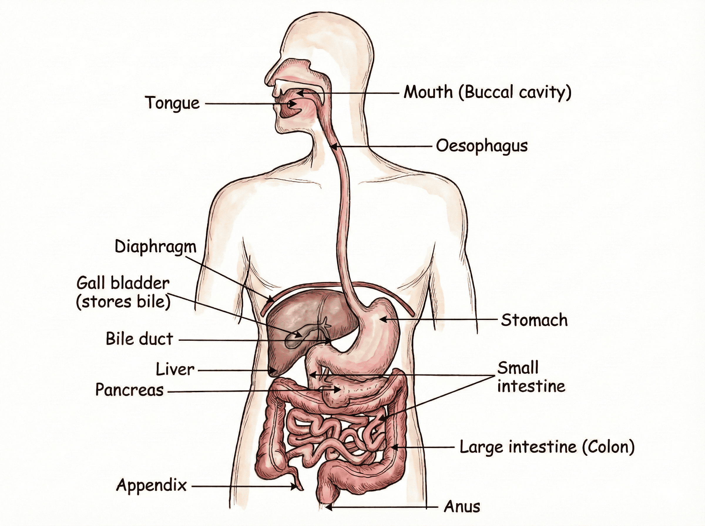
B. Nutrition in Humans (Detailed)
Figure 1.2: Human Digestive System
Alimentary Canal (9 meters long tube):
| Organ |
Secretions |
Action/Function |
| Mouth |
Saliva (Salivary Amylase) |
• Teeth: Chew/Grind.
• Tongue: Tasting/Rolling.
• Amylase: Starch (Complex) → Sugar/Maltose.
|
| Oesophagus |
No Enzymes |
Peristalsis: Rhythmic contraction and relaxation of muscles to push food
down.
|
Stomach
(J-shaped) |
Gastric Glands secrete:
1. HCl
2. Pepsin
3. Mucus |
• HCl: Acidic medium (pH ~1.8) for Pepsin; Kills bacteria.
• Pepsin: Digests Proteins (Partial).
• Mucus: Protects inner lining from acid (prevents ulcers).
|
Small Intestine
(Longest part) |
Receives secretions from Liver & Pancreas |
Site of Complete Digestion.
• Liver (Bile Juice): 1. Makes medium Alkaline. 2.
Emulsification (Large fat globules → Small globules).
• Pancreas (Pancreatic Juice):
- Trypsin: Proteins → Amino Acids.
- Lipase: Emulsified Fats → Fatty Acids + Glycerol.
- Amylase: Remaining Starch → Glucose.
• Intestinal Juice (Walls): Final digestion of all food into simplest forms.
|
| Large Intestine |
No Digestive Enzymes |
Absorbs water and salts from the remaining undigested food. The remaining matter (faeces) is expelled through the anus. |
Dental Caries (Tooth Decay):
Bacteria acting on sugars produce acids that soften or demineralise the enamel. Mass of
bacterial cells together with food particles stick to the teeth to form Dental Plaque.
Saliva cannot reach the tooth surface to neutralize the acid as plaque covers the teeth.
Absorption in Small Intestine:
Inner lining has millions of finger-like projections called Villi. They increase
surface
area for absorption and are richly supplied with blood vessels to take food to every cell.
Why is the Small Intestine longer in herbivores?
Herbivores eat grass containing cellulose, which takes a long time to digest (needs
symbiotic bacteria). Carnivores eat meat, which is easier to digest, so they have shorter intestines.
Pancreas — Dual Role (Digestive + Endocrine):
• Digestive role: Secretes Pancreatic Juice (Trypsin, Lipase, Amylase) into the small intestine.
• Endocrine role: Secretes Insulin (hormone) directly into the blood to regulate blood sugar level.
• Deficiency of Insulin → Diabetes Mellitus (blood glucose level remains high).
2. Respiration (Energy Release)
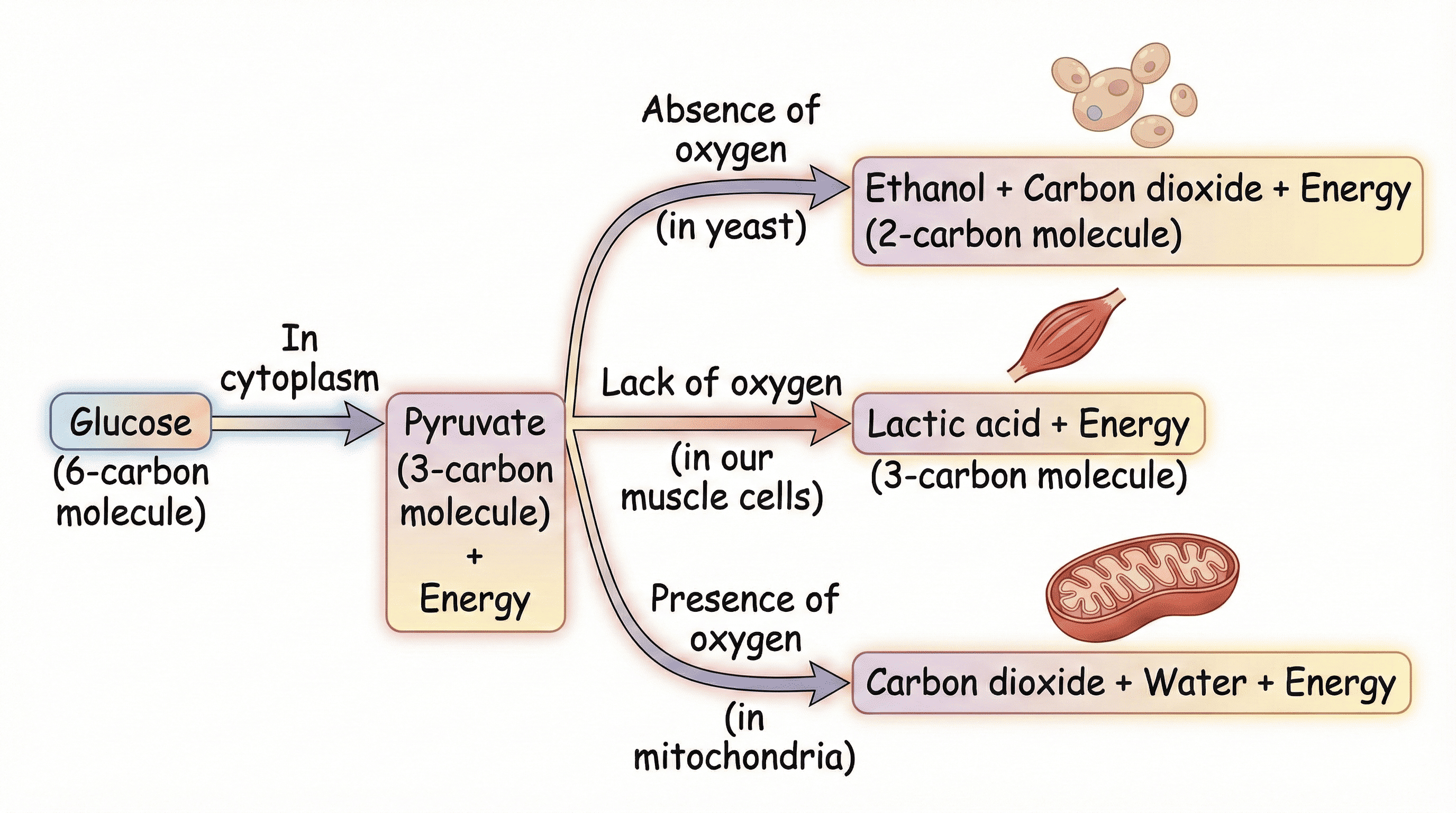
A. Glucose Breakdown Pathways (The Most Repeated PYQ)
Figure 1.2b: Breakdown of Glucose by Various Pathways
Key Exam Point (Muscle Cramps):
When there is a lack of oxygen in muscle cells (sudden activity), Pyruvate converts to
Lactic Acid. Accumulation of lactic acid causes muscle cramps.
Glycolysis — The First Step of Respiration (Common to ALL):
• Location: Cytoplasm (no oxygen needed).
• Process: Glucose (6C) is broken down into Pyruvate (3C) + small amount of Energy.
• After glycolysis, Pyruvate can go 3 ways (depending on oxygen availability):
— Aerobic (in Mitochondria): Pyruvate → CO2 + H2O + Large Energy
— Anaerobic in Yeast (Cytoplasm): Pyruvate → Ethanol (Alcohol) + CO2 + Small Energy
— Anaerobic in Muscles (Cytoplasm): Pyruvate → Lactic Acid + Small Energy
ATP (Adenosine Triphosphate) — The Energy Currency:
• Energy released during respiration is used to synthesize ATP from ADP + inorganic phosphate.
• When energy is needed, ATP is broken down: ATP → ADP + P + Energy (30.5 kJ/mol) per bond.
• ATP is the immediate energy currency of the cell — used for all cellular activities (muscle contraction, protein synthesis, active transport, etc.).
• Full Aerobic Respiration Equation:
C6H12O6 + 6O2 → 6CO2 + 6H2O + Energy (ATP)
| Feature |
Aerobic Respiration |
Anaerobic Respiration |
| Presence of Oxygen |
Takes place in the presence of oxygen. |
Takes place in the absence of oxygen. |
| Site |
Mitochondria. |
Cytoplasm. |
| End Products |
CO2 and H2O. |
Alcohol/Lactic Acid and CO2. |
| Energy Released |
More energy. |
Less energy. |
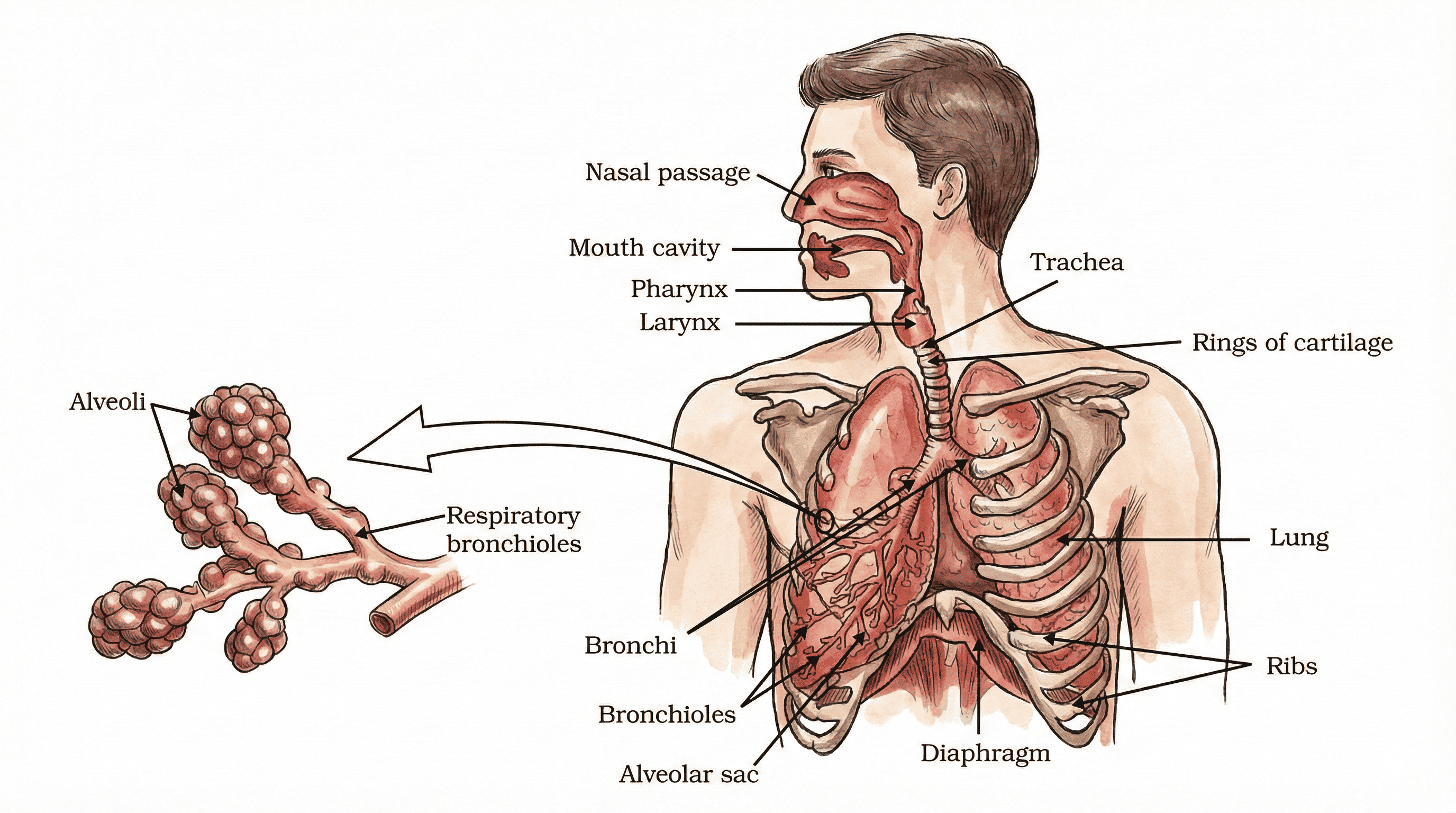
B. Human Respiratory System
Figure 1.3: Human Respiratory System
- Why Trachea has Cartilaginous Rings? Rings of cartilage prevent the trachea (air passage) from collapsing when there is less air inside, ensuring airway always remains open.
- Role of Alveoli (Design for Maximum Efficiency):
— Balloon-like structures that inflate during inhalation.
— Provide a massive surface area (~80 m²) — equivalent to half a tennis court!
— Have extremely thin walls (one cell thick) to allow rapid diffusion of gases.
— Surrounded by an extensive network of blood capillaries for efficient gas exchange.
- Path of Air: Nostrils → Nasal Passage → Pharynx → Larynx → Trachea → Bronchi → Bronchioles → Alveoli (gas exchange).
Mechanism of Breathing:
1. Inhalation: Ribs lift up, Diaphragm flattens → Chest cavity expands → Air rushes
in.
2. Exhalation: Ribs move down, Diaphragm domes up → Chest cavity contracts → Air pushed
out.
*Exchange of gases takes place in Alveoli via diffusion.
Aquatic vs Terrestrial Organisms (Breathing Rate):
Aquatic animals (like fish) breathe faster than terrestrial animals because the amount
of
dissolved oxygen in water is fairly low compared to the amount of oxygen in the air.
Respiratory Pigment:
In humans, Haemoglobin (present in RBCs) carries oxygen from lungs to tissues. It has a
high affinity for oxygen. (CO2 is more soluble in water and is mostly transported in
dissolved
form).
Respiration in Plants:
• Plants exchange gases through stomata (leaves) and lenticels (stems/bark) by diffusion.
• During the day: Both photosynthesis AND respiration occur. The O2 produced by photosynthesis is used in respiration and vice versa — net exchange depends on light intensity.
• During the night: Only respiration occurs → plants take in O2 and release CO2.
• Each part of the plant (roots, stem, leaves) can independently exchange gases with its immediate surroundings.
3. Transportation (Circulation)
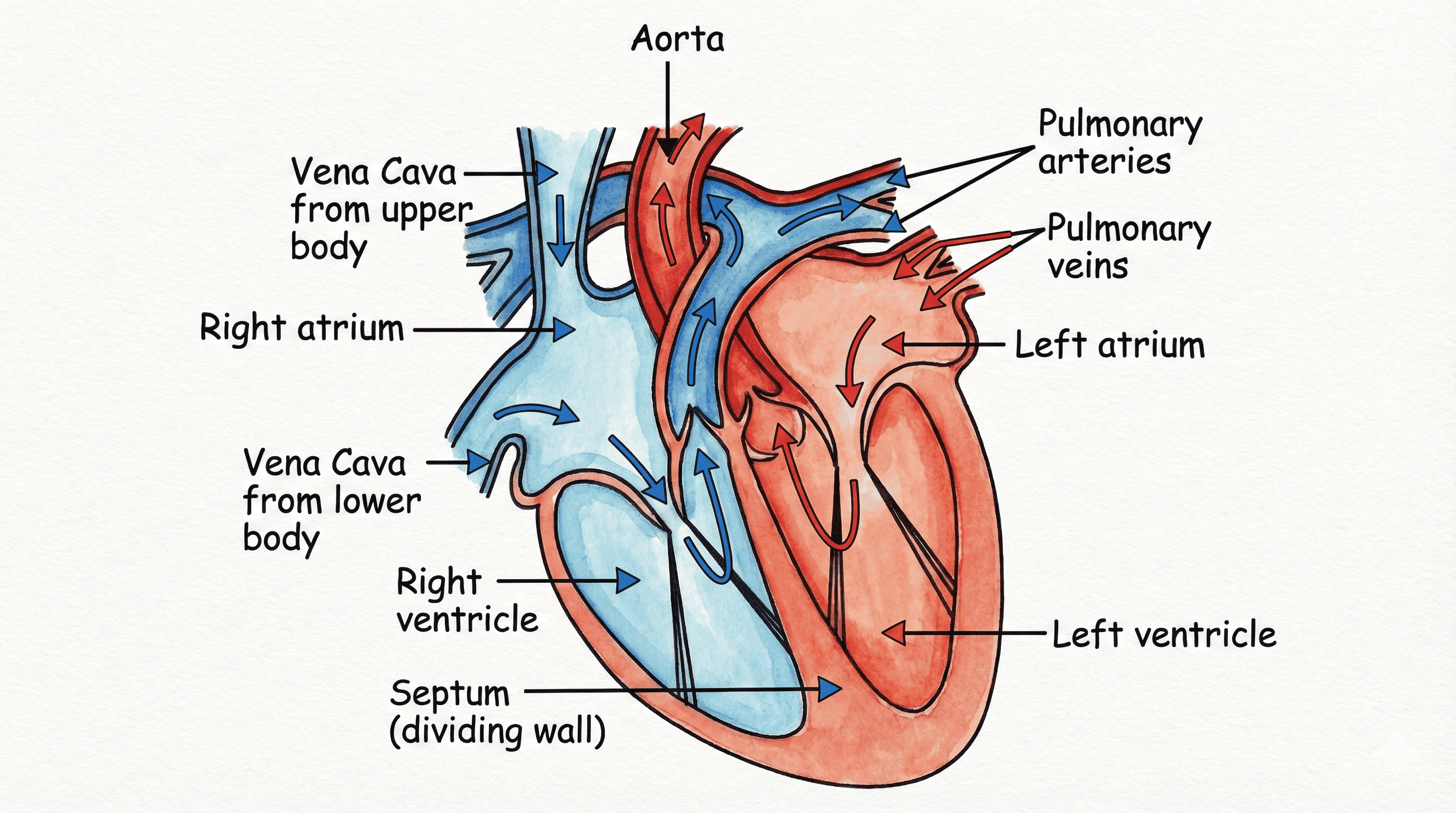
A. Human Heart (Double Circulation)
Figure 1.4: Sectional View of Human Heart
Key Facts About the Human Heart:
• Size: Roughly the size of a fist.
• Has 4 chambers: 2 Atria (upper, thin-walled) + 2 Ventricles (lower, thick-walled).
• Valves between atria and ventricles ensure unidirectional (one-way) blood flow — they prevent backflow when ventricles contract.
• The heart muscle (cardiac muscle) contracts and relaxes rhythmically throughout life without fatigue.
• Oxygenated and Deoxygenated blood travel in completely separate circuits — they never mix.
Figure 1.4b: Schematic of Double Circulation
Double Circulation Explained:
Blood passes through the heart twice in one complete cycle.
1. Pulmonary Circulation: Blood moves from Heart → Lungs → Heart (Oxygenation).
2. Systemic Circulation: Blood moves from Heart → Body Organs → Heart (Supply O2).
Step-by-Step Blood Flow:
- Vena Cava brings CO2-rich blood from body to Right
Atrium.
- Right Atrium relaxes (fills) → Contracts → Blood goes to Right Ventricle.
- Right Ventricle pumps blood to Lungs via Pulmonary Artery (for oxygenation).
- Pulmonary Veins bring O2-rich blood from Lungs to Left
Atrium.
- Left Atrium contracts → Blood goes to Left Ventricle.
- Left Ventricle (Thickest wall) pumps blood to body via Aorta.
(Valves ensure unidirectional flow).
Why is separation of Oxygenated and Deoxygenated blood necessary?
Separation allows for a highly efficient supply of oxygen to the body at all times. This is especially important for birds and mammals (warm-blooded/endothermic animals), which constantly use energy to maintain their body temperature. A mixed supply would reduce efficiency.
Blood Pressure (BP):
• Blood pressure is the pressure exerted by blood against the walls of blood vessels.
• Normal BP = 120/80 mmHg (Systolic/Diastolic). Measured with a Sphygmomanometer.
• Systolic pressure (120): Pressure when ventricles contract (pumping).
• Diastolic pressure (80): Pressure when ventricles relax (filling).
• Hypertension (High BP): If blood pressure is consistently high, it can cause damage to blood vessels and lead to heart disease or stroke.
Why do Ventricles have thicker walls than Atria?
Ventricles have to pump blood into various organs (Left V → Body; Right V → Lungs), so they need thicker muscular walls to generate high pressure. The Left Ventricle has the thickest wall as it must pump blood to the entire body.
Circulation in Different Animals:
• Fish (2-chambered heart: 1 Atrium + 1 Ventricle): Single Circulation — blood passes through the heart once per cycle. Deoxygenated blood → Heart → Gills (oxygenated) → Body → Heart.
• Amphibians & Reptiles (3-chambered heart): Incomplete Double Circulation — oxygenated and deoxygenated blood mix in the single ventricle.
• Mammals & Birds (4-chambered heart): Complete Double Circulation — complete separation prevents mixing, giving maximum efficiency.
Components of Blood:
| Component |
Description & Function |
| Plasma |
Liquid part (~55%). Transports food, CO2, and metabolic wastes in dissolved form. |
| RBC (Red Blood Cells) |
Contains Haemoglobin. Transports oxygen from lungs to body tissues. |
| WBC (White Blood Cells) |
Fight infections; part of the immune system. |
| Platelets |
Help in clotting of blood at points of injury to plug leaks and prevent loss of pressure in the pumping system. |
Arteries vs. Veins vs. Capillaries:
| Feature |
Arteries |
Veins |
Capillaries |
| Direction |
Away from heart |
Towards heart |
Connect arteries & veins |
| Blood Pressure |
High |
Low |
Very low |
| Wall Thickness |
Thick & elastic |
Thin |
One cell thick |
| Valves |
No valves |
Have valves (prevent backflow) |
No valves |
| Blood Type |
Oxygenated (except Pulmonary Artery) |
Deoxygenated (except Pulmonary Vein) |
Mixed / Exchange site |
B. Transportation in Plants
| Xylem |
Phloem |
| Transports Water & Minerals. |
Transports Food (Sucrose, Amino Acids). |
| Direction: Unidirectional (Upwards only). |
Direction: Bidirectional (Source → Sink). |
| Physical/Passive Process: Transpiration Pull (Suction). |
Active Process: Translocation (Uses Energy/ATP). |
How Water Rises in Xylem — Two Forces:
1. Root Pressure: Osmosis pushes water from soil into root cells and then into Xylem. More significant at night when transpiration is minimal. Evidence: Guttation (water drops on leaf edges at night).
2. Transpiration Pull: Evaporation of water from leaves creates a suction (pull) that draws water upward. This is the major force during the day and in tall trees.
Translocation in Phloem (Detailed NCERT Mechanism):
1. Food (sucrose) made in leaves is actively loaded into the sieve tubes of phloem using ATP (energy).
2. This increases the osmotic pressure inside phloem → water moves in from nearby xylem by osmosis.
3. This creates high pressure in the phloem, which moves the food from the source (leaves) to areas of lower pressure (sink) — roots, fruits, growing tips.
*This is why phloem transport is bidirectional — it goes wherever the sink is.
Why is Transport in Plants Slower than in Animals?
• Plants do not move; they have a large proportion of dead cells in many tissues (e.g., xylem).
• Their energy needs are therefore low.
• They can use relatively slow, physical transport systems (transpiration pull, osmosis).
• Animals need fast transport due to high activity and energy demands.
4. Excretion
Definition of Excretion:
The biological process of removing toxic metabolic wastes from the body is called Excretion.
• In humans, the main waste is nitrogenous waste produced from the breakdown of proteins and excess amino acids — primarily Urea (also Uric Acid, Ammonia).
• Other wastes include CO2 (from respiration) and excess water and salts.
• Why must nitrogenous waste be removed? Urea and ammonia are toxic to cells — their accumulation can damage organs and disrupt metabolism.
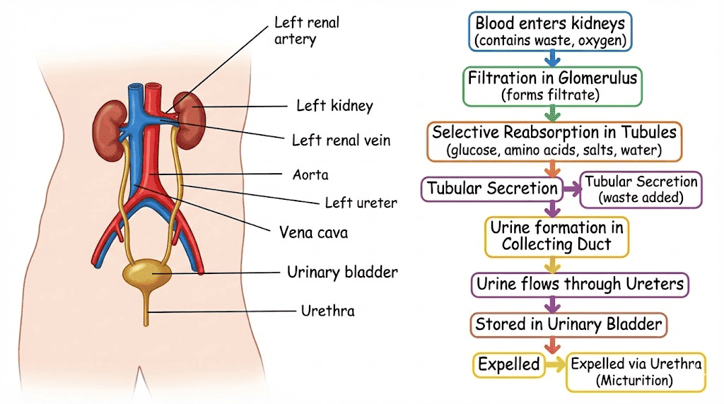
A. Human Excretory System
Figure 1.5: Human Excretory System
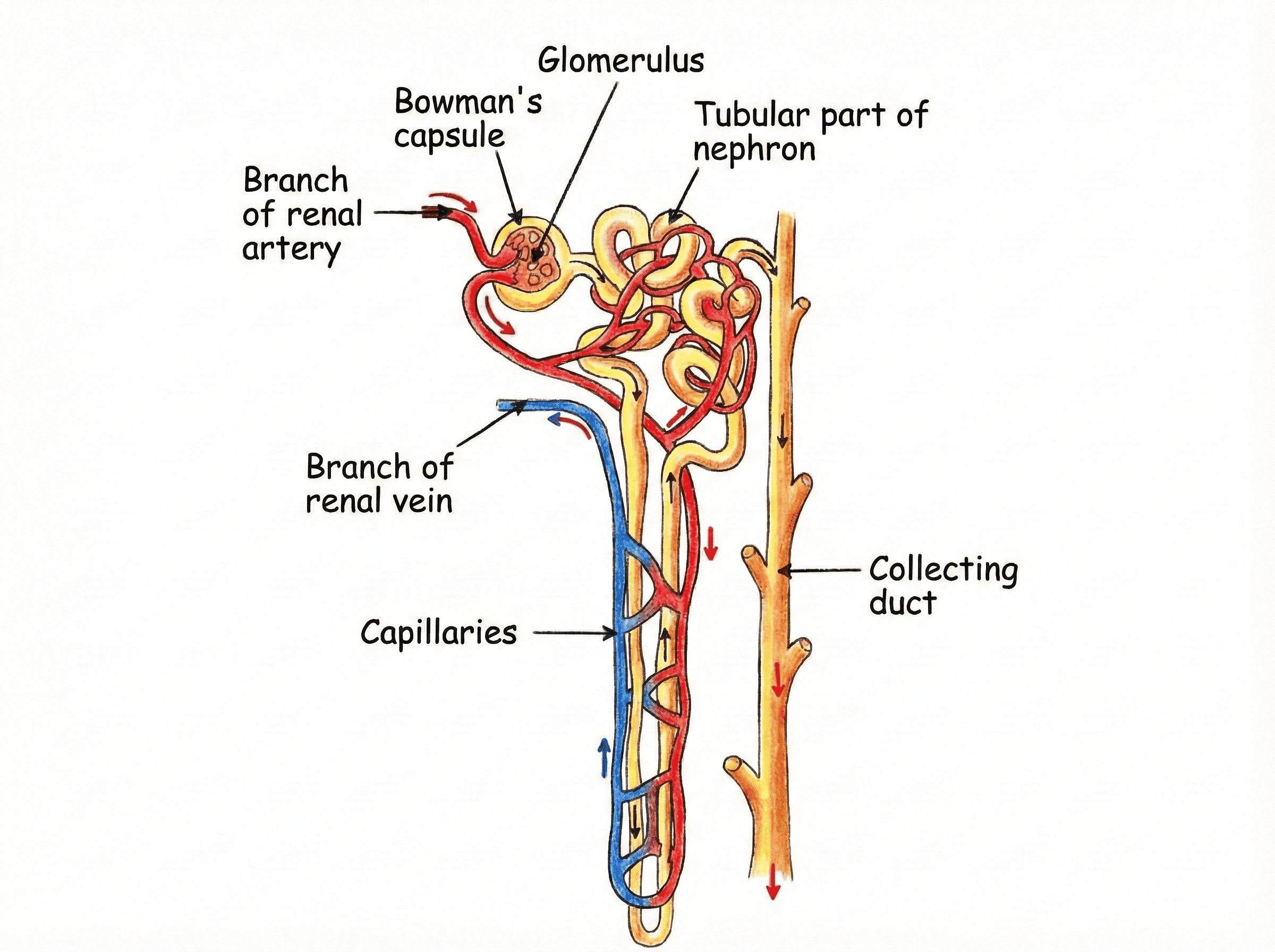
Figure 1.6: Structure of Nephron
Nephron: Filtration unit of Kidney.
Formatting Urine:
1. Filtration: Blood filtered in Glomerulus (Bowman's Capsule).
2. Reabsorption: Useful substances (Glucose, Amino acids, Salts, Water) reabsorbed in
tubule.
3. Secretion: Waste remains as Urine → Collecting Duct → Ureter → Urinary Bladder →
Urethra.
Hemodialysis (Artificial Kidney):
Used in kidney failure. Filters blood using dialysing fluid (same osmotic pressure as blood but NO
nitrogenous
waste). Difference: No Reabsorption occurs in dialysis.
B. Excretion in Plants
Plants produce far less nitrogenous waste than animals (since they don't have complex movement/metabolism). They use a variety of strategies:
- Oxygen: Released via stomata and lenticels (by-product of photosynthesis).
- CO2 & Water Vapour: Released via stomata (by-product of respiration).
- Transpiration: Excess water lost through stomata (leaves) and lenticels (stem/bark).
- Vacuoles: Many waste products are stored in the cell vacuoles (e.g., tannins, alkaloids, essential oils).
- Old Leaves: Wastes accumulated in leaves are shed seasonally (autumn leaf fall).
- Resins & Gums: Some wastes stored in old xylem as resins and gums (e.g., pine resin). Note: Starch is NOT a waste — it is stored food.
- Excretion into Soil: Some waste products are actively excreted into the soil around the roots. These can sometimes inhibit the growth of other nearby plants (allelopathy).
Lymph — The Third Circulatory Fluid:
• Formation: Some plasma, proteins, and white blood cells (lymphocytes) seep out through the pores of capillary walls into the intercellular spaces, forming the tissue fluid (lymph).
• Composition: Similar to plasma but NO Red Blood Cells; contains WBCs (lymphocytes) and proteins.
• Functions:
1. Carries digested and absorbed fat from the intestine to the blood.
2. Drains excess intercellular fluid back into the blood (via lymph vessels → larger veins).
• Note: Lymph vessels open into larger Veins (NOT arteries).
-
Why do veins have valves while arteries do not?
Hint: Veins carry blood under low pressure against gravity (need to
prevent
backflow). Arteries have high pressure from the heart.
-
A plant is kept in a dark room. Will it carry out Photosynthesis or Respiration?
Hint: It will perform Respiration (takes in O2, gives out CO2).
Photosynthesis needs Light.
-
Why is the rate of breathing in aquatic organisms much faster than in terrestrial organisms?
Hint: Dissolved Oxygen in water is very low compared to O2 in the
air.
🧪 NCERT EXPERIMENT ZONE (Life Processes)
1. Chlorophyll is necessary for Photosynthesis:
•
Plant: Variegated leaf (e.g., Croton/Money Plant - patches of green and
white).
•
Process: Keep in dark (destarch) → Sunlight 6 hrs → Boil in alcohol (remove
chlorophyll) → Iodine test.
•
Observation: Only green parts turn
Blue-Black (Starch present). White
parts (no chlorophyll) do not.
Figure 1.E1: Chlorophyll is needed for photosynthesis
2. CO₂ is necessary for Photosynthesis:
•
Plant: Two destarched potted plants.
•
Setup: Bell jars. One with Potassium Hydroxide (KOH) in a watch glass.
•
Role of KOH: Absorbs CO₂.
•
Observation: Leaf from jar with KOH does
NOT turn Blue-Black with
Iodine.
Figure 1.E2: CO2 is needed for photosynthesis
(a) with potassium
hydroxide (b)
without potassium hydroxide
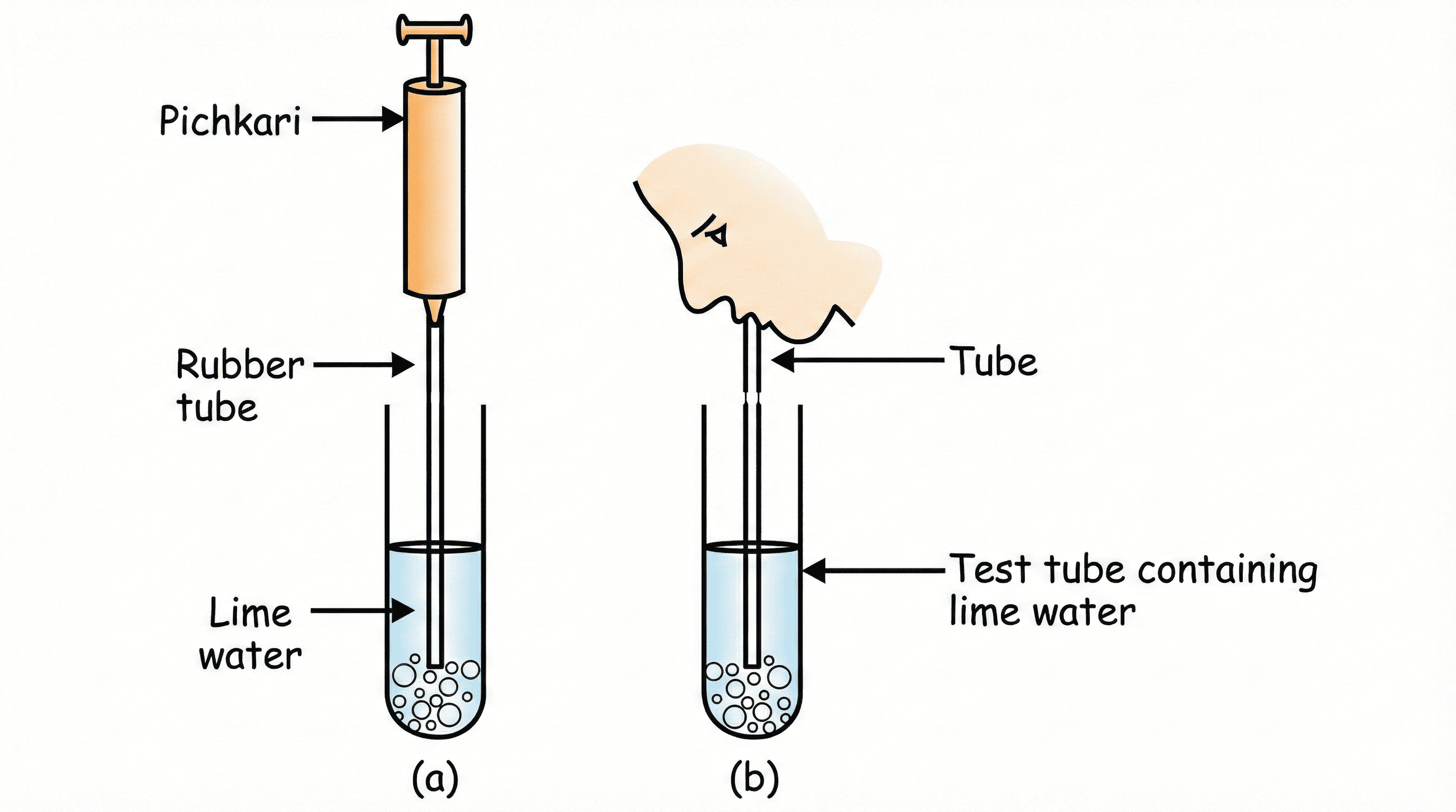
3. CO₂ is released during Respiration:
•
Test: Blow air into Lime Water.
•
Observation: Lime Water turns
Milky.
•
Conclusion: Exhaled air contains Carbon Dioxide.
Figure 1.E3: CO2 is released during Respiration
4. Action of Saliva on Starch:
•
Test Tube A: Starch + Saliva.
Test Tube B: Starch only.
•
Result: After 20 mins, Tube A does NOT turn blue-black (Starch digested by Amylase).
Tube
B turns blue-black.

.png)
 vs Closed (Flaccid).png)
.png)
 before starch test (b) after starch test.png)
 with potassium hydroxide (b) without potassium hydroxide.png)
 VARDAAN COMET